

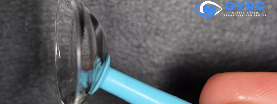
Scleral Lenses for Sjogren’s Syndrome: Patient Guide
Scleral lenses may help selected people with Sjogren’s syndrome whose dry eye, ocular surface disease, or unstable vision is not adequately managed with conventional correction alone. A scleral lens vaults over the cornea and holds sterile, preservative-free saline in the space beneath the lens during wear. It may support comfort, protect the corneal surface, and create a more stable optical surface, but it does not cure Sjogren’s disease or replace medical dry-eye care.
Candidacy depends on the ocular surface, eyelids, tear film, corneal health, vision, handling ability, previous treatments, and how the eye responds to a diagnostic lens. Dr. Edward Boshnick evaluates those factors together rather than recommending a scleral lens from the diagnosis alone.
Key Takeaways
- Sjogren’s disease can cause severe aqueous-deficient dry eye, while eyelid and meibomian-gland problems may add an evaporative component.
- A scleral lens can maintain a fluid reservoir over the cornea during wear and may also correct vision through its front optical surface.
- The lens treats neither the systemic autoimmune disease nor every cause of eye pain, redness, light sensitivity, or blurred vision.
- Front-surface dryness, fogging, bubbles, deposits, handling difficulty, or an unhealthy corneal response may limit wear and require changes.
- Safe use requires an individualized design, prescribed solutions, water avoidance, scheduled follow-up, and prompt evaluation of warning symptoms.
How Sjogren’s Disease Can Affect the Eyes
Sjogren’s is an autoimmune disease that can affect glands and other tissues throughout the body. In the eyes, reduced tear secretion and inflammation can destabilize the tear film and damage the ocular surface. Common symptoms include burning, grittiness, light sensitivity, fluctuating blur, mucus, redness, eye fatigue, and difficulty reading or using screens. Some people also have dry mouth, fatigue, joint symptoms, or another autoimmune condition.
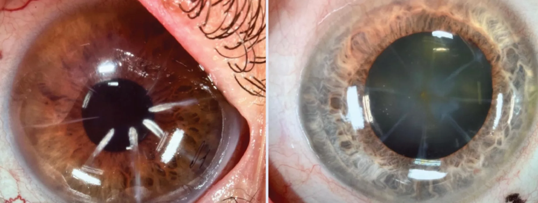
Dry eye is not one uniform problem. A person with Sjogren’s may have marked aqueous tear deficiency as well as blepharitis or meibomian-gland dysfunction that increases evaporation. Corneal nerve changes and neuropathic pain can also affect symptoms. The intensity of symptoms does not always match the amount of staining or other examination findings, so both the patient’s experience and objective findings matter.
How a Scleral Lens Works
A scleral lens is a large-diameter, oxygen-permeable rigid lens. It rests on the conjunctival tissue over the sclera rather than bearing on the central cornea. Before insertion, the bowl is filled completely with sterile, preservative-free saline recommended for that patient. The lens then vaults over the cornea and maintains a fluid reservoir beneath it during wear.
The fluid reservoir may reduce direct exposure and eyelid friction over the cornea. The smooth front surface of the lens can also improve optical quality when an unstable tear film or irregular corneal surface contributes to fluctuating or distorted vision. These effects occur while the lens is worn; they do not turn the lens into a cure for the autoimmune disease.
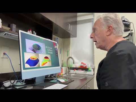
This original Eye Freedom video is educational. One patient’s experience cannot predict another patient’s comfort, vision, or wearing time.
What Scleral Lenses May Help With
- Ocular surface support during wear: The fluid reservoir may help protect and hydrate the corneal surface for an appropriately fitted patient.
- More stable vision: The lens may reduce optical fluctuation caused by an unstable tear film or irregular corneal surface.
- Reduced mechanical exposure: The vault may shield the cornea from some eyelid friction and environmental exposure.
- An alternative to standard contact lenses: A selected patient who cannot tolerate a conventional soft or corneal gas-permeable lens may be evaluated for a scleral design.
Published studies and reviews support scleral lenses as an option for selected moderate-to-severe ocular surface disease, but the evidence includes retrospective studies, case series, mixed diagnoses, and different lens designs. Average study results do not guarantee a particular outcome for one patient.
What a Scleral Lens Cannot Do
A scleral lens does not eliminate Sjogren’s disease, restore normal tear-gland function, treat every source of ocular inflammation, or replace care from the professionals managing the systemic condition. It also cannot guarantee all-day wear, normal vision, or complete relief from burning, pain, light sensitivity, or dryness.
Some patients continue prescribed drops, ointments, eyelid therapy, serum tears, punctal treatment, moisture protection, or systemic care. Timing medications around lens wear must be discussed with the prescribing professional. Do not place a medication beneath a scleral lens unless specifically directed.
For broader medical and ocular-surface management, see the guide to dry eye syndrome and meibomian-gland dysfunction.
Who May Be Evaluated for Scleral Lenses?
An evaluation may be reasonable when severe dryness, surface exposure, contact-lens intolerance, or fluctuating vision continues despite appropriate care, or when a diagnostic lens may clarify how much vision is optically correctable. The goal may be ocular-surface support, optical rehabilitation, or both.
Active infection, uncontrolled inflammation, an epithelial defect, significant eyelid or conjunctival disease, reduced corneal sensation, inability to handle the lens safely, poor hygiene, or an unacceptable corneal response may require treatment first or may limit candidacy. Other severe ocular-surface disorders, including graft-versus-host disease, Stevens-Johnson syndrome, and neurotrophic keratitis, require their own individualized medical and lens planning; read the focused severe ocular surface disease guide.
How Dr. Boshnick Evaluates Sjogren’s Dry Eye
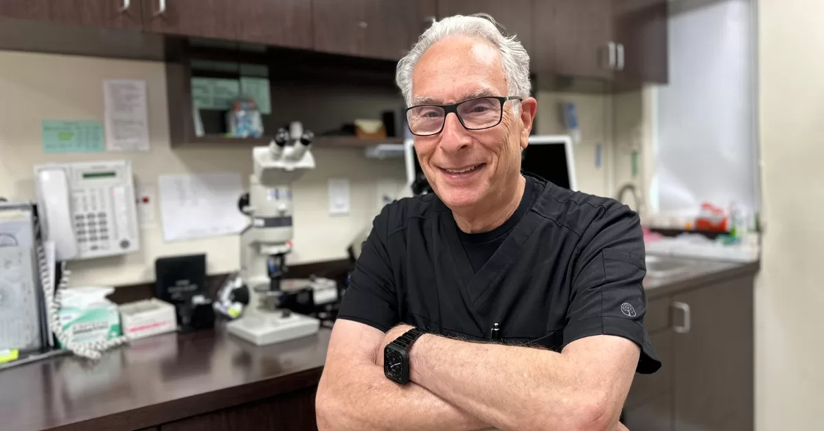
Dr. Boshnick reviews symptoms, systemic diagnoses, medications, previous dry-eye care, contact-lens history, visual demands, and the patient’s goals. The examination considers the cornea, conjunctiva, eyelids, tear film, staining, vision, refraction, and relevant surgical or medical history.
Depending on the eye, testing may include corneal topography, tomography, ocular-surface imaging, or a diagnostic scleral-lens trial. During the trial, he evaluates clearance over the cornea and limbus, the landing relationship, centration, movement, bubbles, comfort, vision, and changes as the lens settles. A visually sharp lens is not acceptable if the fit or physiological response is unhealthy.
What Happens During a Custom Scleral Lens Fitting?
- History and examination: The ocular surface, tear film, eyelids, vision, prior treatments, handling needs, and medical context are reviewed.
- Measurements and diagnostic lens: Corneal shape and other measurements guide an initial lens. After application, the fit and visual response are assessed as the lens settles.
- Design refinement: Diameter, sagittal depth, limbal clearance, landing geometry, optical power, material, and surface treatment may be adjusted. Some eyes may be evaluated for technologies such as EyePrintPRO or wavefront-guided optics when clinically appropriate.
- Handling and care training: The patient learns filling, insertion, bubble checks, removal, cleaning, disinfection, storage, and what to do if symptoms develop.
- Follow-up: Vision, comfort, wearing time, fit, surface response, and lens condition are reviewed. More than one design change may be needed.
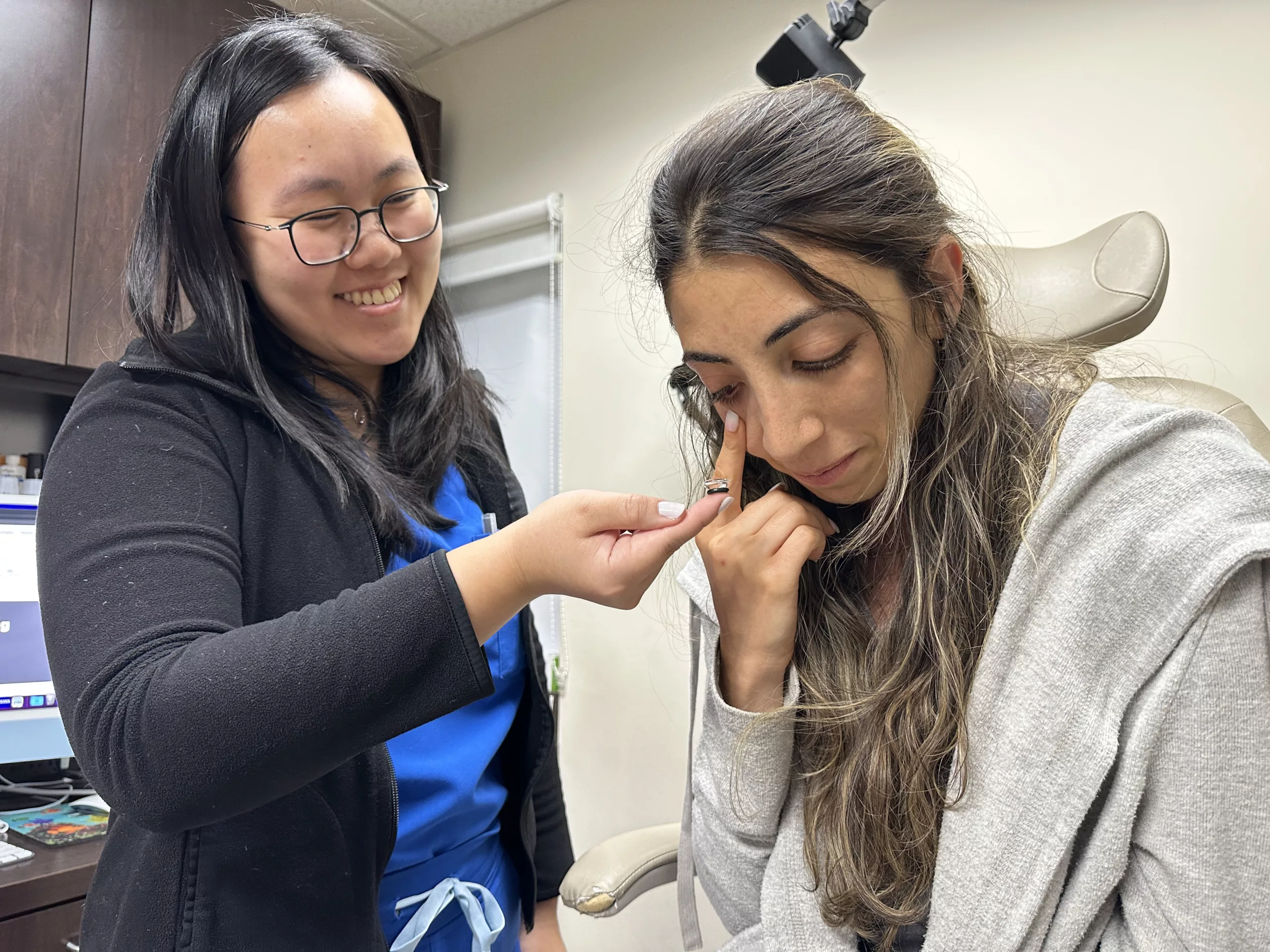

This original Eye Freedom video shows lens handling in a clinical context. Patients receive individualized instruction for their own lenses and eyes.
Learn more about why custom fitting and follow-up matter.
Front-Surface Dryness, Wetting, and Midday Fogging
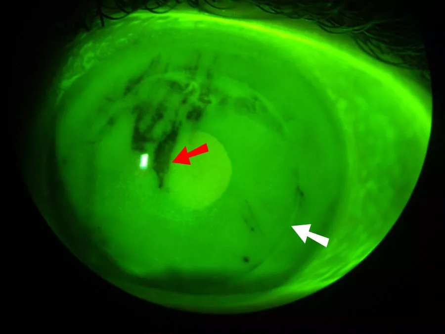
The fluid reservoir is beneath the lens, but the front surface is exposed to the eyelids, tears, air, and deposits. A patient can therefore have a well-filled reservoir and still experience front-surface dryness, poor wetting, blur, or discomfort. Lens material, surface condition, tear-film quality, eyelids, products, environment, and wear time can all contribute.
Midday fogging is blur caused by debris or turbidity in or around the post-lens reservoir. Inflammation, tear debris, excessive or uneven clearance, landing-zone alignment, bubbles, filling technique, and solution compatibility may contribute. Removing, cleaning, refilling, and reapplying the lens may temporarily clear the view, but repeated fogging deserves evaluation rather than assuming it is normal.
Do not change filling solutions, add medication under the lens, polish the lens, or extend wear based only on online advice. Dr. Boshnick may adjust the lens design, surface treatment, care products, application technique, or broader ocular-surface plan after identifying the likely cause.
Cleaning, Disinfection, and Water Safety
Sterile saline used to fill a scleral lens is not necessarily a disinfectant. Reusable lenses must be cleaned, disinfected, rinsed, and stored with the specific system prescribed for the lens material and patient. Wash and dry hands before handling, use fresh products as directed, and replace the case and lenses on the recommended schedule.
- Never use tap, bottled, distilled, or homemade water to fill, rinse, clean, or store a contact lens.
- Remove lenses before showering, swimming, or using a hot tub unless an eye-care professional provides different emergency instructions.
- Do not sleep in scleral lenses unless the treating professional has prescribed a specific medically supervised protocol.
- Do not top off old storage solution or substitute a product that was not approved for the lens and eye.
- Keep current backup glasses available in case lenses must be removed.
Wear Time, Replacement, and Follow-Up
There is no single safe wearing time or replacement interval for every person with Sjogren’s disease. Initial wear may be gradual. The schedule depends on surface health, comfort, vision, lens fit, fogging, deposits, medications, material, handling, and the response observed at follow-up.
A lens may need replacement when the prescription or eye changes, the fit is no longer appropriate, the surface is damaged or poorly wetting, deposits cannot be managed, or the lens becomes warped, cracked, or chipped. Follow-up is usually more frequent during fitting and is then individualized. New symptoms should not wait for the next routine visit.
When to Remove a Lens and Seek Prompt Care
Remove the lens and contact an eye-care professional promptly for increasing pain, marked redness, light sensitivity, discharge, sudden or persistent loss of vision, excessive tearing, a new white spot on the cornea, or symptoms that continue after removal. Contact-lens-related microbial keratitis can threaten vision and should not be managed by repeatedly reinserting the lens.
Patients with severe ocular-surface disease, reduced corneal sensation, immune suppression, or a history of epithelial breakdown may need additional precautions and coordination with a corneal specialist or another physician.
Discussing Scleral Lenses With Dr. Boshnick
Bring a medication list, prior dry-eye and autoimmune diagnoses, current glasses and contact lenses, previous imaging or corneal records, and a summary of treatments already tried. It is useful to describe when symptoms are worst, how vision changes during the day, and which activities are limited.
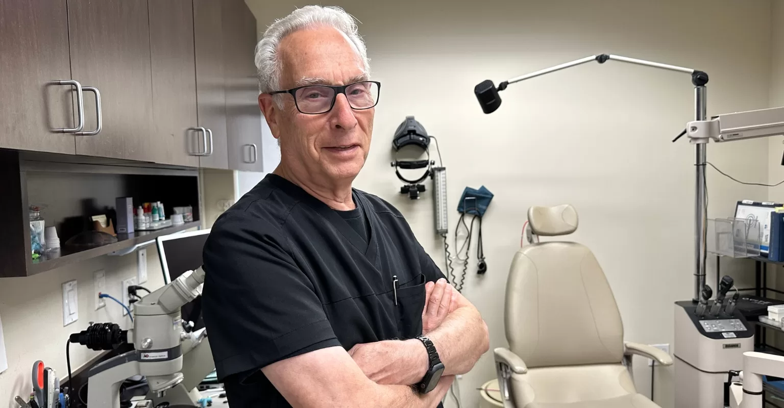
To discuss whether a diagnostic scleral-lens evaluation is appropriate, request an evaluation with Dr. Edward Boshnick or call 305-271-8206. The practice evaluates patients who travel from across the United States and internationally.
Frequently Asked Questions
Can people with Sjogren’s syndrome wear contact lenses?
Some can, but the diagnosis alone does not determine candidacy. Severe tear deficiency, inflammation, eyelid disease, corneal staining, reduced sensation, handling ability, and prior lens intolerance all matter. A custom scleral lens may be considered when a conventional lens is not tolerated, but only after an eye-health and diagnostic-lens evaluation.
How may scleral lenses help Sjogren’s dry eye?
A scleral lens vaults the cornea and maintains sterile, preservative-free saline beneath it during wear. This may support the ocular surface and improve optical stability for selected patients. Results vary, and the lens does not treat the underlying autoimmune disease.
Do scleral lenses cure dry eye caused by Sjogren’s disease?
No. They may support comfort, corneal protection, or vision during wear, but they do not cure Sjogren’s disease or restore normal tear production. Medical dry-eye and systemic care may still be needed.
Are scleral lenses always comfortable for severe dry eye?
No. Many selected patients seek them because the lens vaults the sensitive cornea, but comfort depends on fit, landing alignment, surface wetting, deposits, eyelid interaction, inflammation, handling, and the individual eye. Discomfort should be evaluated rather than accepted as unavoidable.
Why can a scleral lens become dry or foggy?
The front surface can dry because it remains exposed to the tear film and environment. Fogging may involve tear debris, inflammation, lens clearance, landing alignment, bubbles, products, or application technique. Recurrent blur needs examination so the likely cause can be addressed.
How long can someone with Sjogren’s wear scleral lenses each day?
Wear time is individualized. It depends on ocular-surface health, comfort, vision, lens fit, fogging, deposits, and the response seen at follow-up. Patients should follow the prescribed schedule and remove the lens if warning symptoms occur.
Can prescription dry-eye drops still be used with scleral lenses?
Often they remain part of care, but timing and product compatibility must be coordinated with the prescribing professional. Do not place medication in the lens reservoir unless specifically instructed to do so.
What happens during a scleral-lens fitting for Sjogren’s disease?
The process includes an eye-health and tear-film assessment, corneal measurements, a diagnostic lens, evaluation after settling, vision testing, design refinement, handling instruction, and follow-up. More than one visit or lens change may be necessary.
When do symptoms require urgent evaluation?
Increasing pain, pronounced redness, light sensitivity, discharge, sudden or persistent blur, excessive tearing, or a corneal white spot requires prompt professional guidance. Remove the lens and do not resume wear until advised.
Medical Sources
- Sjogren’s Foundation: Ocular health, dry eye, and Sjogren’s disease
- American Academy of Ophthalmology EyeWiki: Dry eye in Sjogren’s syndrome
- Peer-reviewed case series: Scleral lenses in the management of ocular surface disease
- Peer-reviewed review: Therapeutic uses, patient selection, and special considerations
- Peer-reviewed review: Scleral lenses for dry eye without corneal irregularity
- Peer-reviewed review: Midday fogging of scleral lenses
- CDC: Cleaning, disinfecting, and storing contact lenses
- CDC: Keeping water away from contact lenses
- CDC: Contact-lens-related eye infections and warning symptoms
This page is educational and does not provide a diagnosis or individualized treatment plan. A specialty examination is required to determine whether scleral lenses are appropriate.
