7800 SW 87 AVE SUITE B-270 MIAMI, FLORIDA 33173

Visante Optical Coherence Tomography: A Comprehensive Tool for Evaluating the Cornea and Anterior Segment

When it comes to diagnosing and managing diseases and conditions related to the front portion of the eye, the Visante Optical Coherence Tomography (OCT) stands out as a critical instrument. It offers in vivo cross-sectional images of ocular structures, providing a wealth of information that aids in detailed analysis and improved understanding of the eye’s anterior segment. This article explains the importance of Visante OCT in the ophthalmological field and the various ways it assists in restoring functional vision and ocular comfort for patients.

What is Visante Optical Coherence Tomography?

Optical Coherence Tomography (OCT) is a non-invasive imaging test that uses light waves to take cross-section pictures of your retina, the light-sensitive tissue lining the back of the eye. The Visante OCT takes this technology a step further by focusing on the anterior segment of the eye, which includes the cornea, anterior chamber, crystalline lens, and iris.

This cutting-edge technology generates detailed, cross-sectional images of these ocular structures, offering a depth of insight unattainable with other technologies. This invaluable tool can be instrumental in diagnosing and managing numerous anterior segment diseases and conditions such as keratoconus, corneal transplants, corneal ectasia, narrow-angle glaucoma, and various corneal dystrophies and degenerations.

The Clinical Applications of Visante OCT

The clinical applications of anterior segment OCT are vast and incredibly beneficial. Our office uses optical coherence tomography to help us gain a better understanding of many complex corneal conditions that we encounter. We are now using OCT to design better specialty contact and scleral lenses for our patients.

One of the primary uses of Visante OCT is in the evaluation of keratoconus, a condition where the cornea becomes irregularly shaped, causing distorted vision. With OCT, we can visualize and monitor the progression of keratoconus in detail, which assists us in selecting the best treatment and management options for our patients.

Visante OCT also plays a crucial role in the assessment and management of corneal transplants. It allows us to examine the corneal layers and graft interface in detail, ensuring the transplant is healthy and functioning correctly.

In cases of narrow-angle glaucoma, Visante OCT helps us evaluate the anterior chamber angle, which is crucial in diagnosing and treating this condition. It provides clear, detailed images of the iris and angle, helping us make accurate assessments.

The technology is not limited to these conditions alone. Its high-resolution imaging capability makes it valuable in diagnosing and managing a variety of corneal dystrophies and degenerations, providing us with an understanding that would be impossible with conventional imaging techniques.

Visante OCT in Action

An excellent example of the practical application of Visante OCT is in the evaluation of a scleral lens over an eye with an advanced case of keratoconus with hydrops. Hydrops is a condition where the most posterior corneal membrane ruptures, allowing fluid from inside the eye to enter the center of the cornea, causing it to become hazy. With Visante OCT, we can clearly see the torn corneal membrane, which is instrumental in monitoring the condition and ensuring the patient maintains clear (20/30), comfortable vision with all-day lens wear.

Conclusion

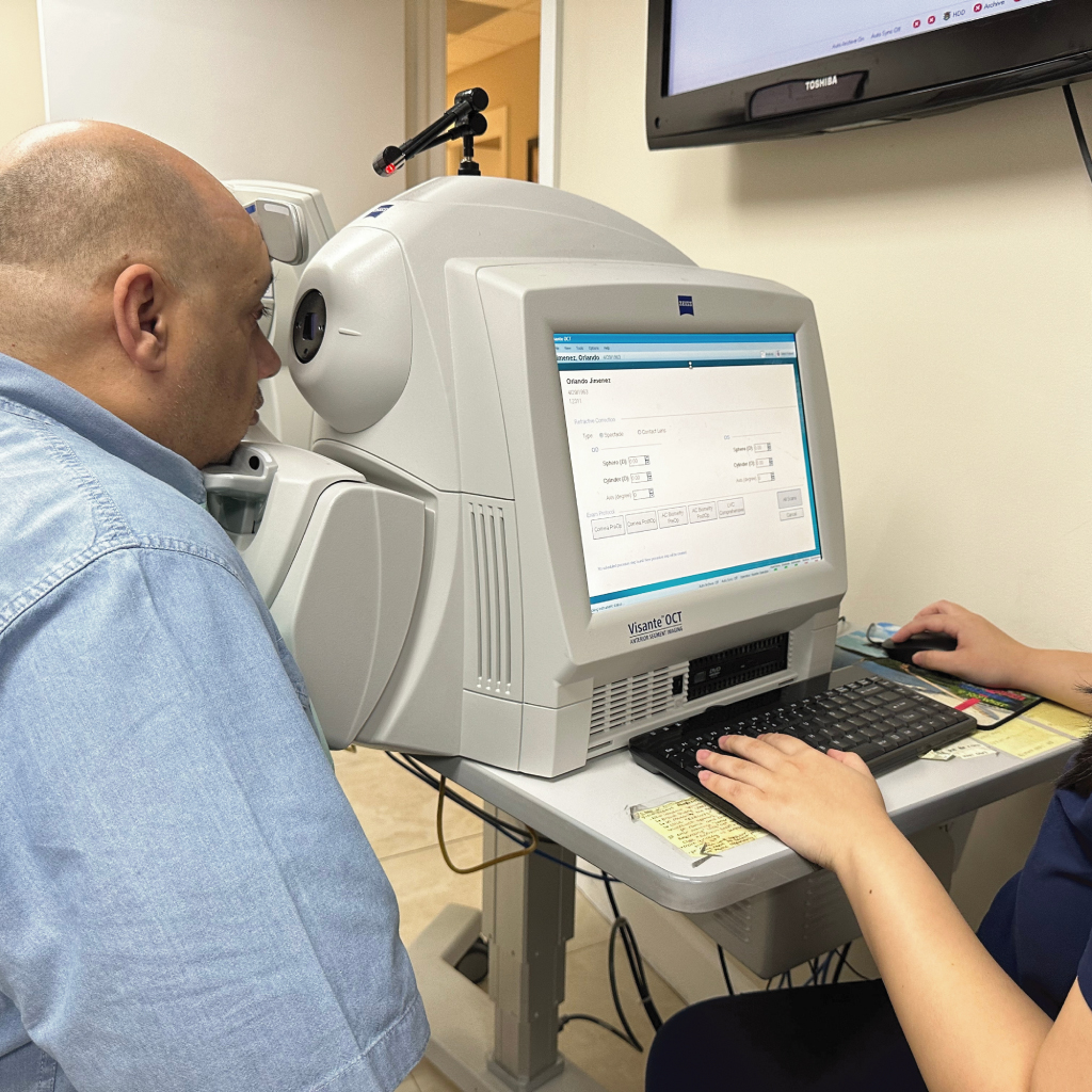
The Visante Optical Coherence Tomography is more than just a tool; it is an essential aspect of modern ophthalmology that enhances our understanding and management of various corneal and anterior segment conditions. By helping us design better specialty contact and scleral lenses, it plays a crucial role in our mission to help our patients regain functional vision and maintain excellent ocular comfort.

By mapping the anterior segment in detail, Visante OCT aids in the creation of personalized treatment plans tailored to each patient’s unique corneal profile. This personalized care approach can lead to more effective treatment outcomes and improved patient satisfaction.

Visante OCT also plays an essential role in monitoring disease progression and treatment efficacy. For example, in conditions like keratoconus, narrow-angle glaucoma, or after corneal transplants, the ability to visualize minute changes over time can be instrumental in determining if a treatment is working or if adjustments need to be made. The technology’s ability to track changes over time provides valuable data, helping us make informed decisions and adjust treatment plans as necessary.

Furthermore, the high-resolution images generated by Visante OCT can be invaluable for patient education. By allowing patients to see detailed images of their eyes, we can better explain their condition and the rationale behind their treatment plan. This can lead to increased patient understanding, trust, and adherence to treatment.

In essence, Visante OCT is a powerful tool in the hands of ophthalmologists and eye care practitioners. It enhances diagnostic accuracy, informs treatment plans, monitors disease progression, and improves patient education – all of which contribute to improved eye health outcomes.

In the ever-evolving field of ophthalmology, the importance of adopting and utilizing advanced technology like Visante OCT cannot be overstated. As we continue to understand and treat a wide variety of corneal and anterior segment conditions, this technology is poised to remain an invaluable tool in our arsenal.

In conclusion, Visante Optical Coherence Tomography, with its detailed cross-sectional imaging, high resolution, and versatile applications, truly is revolutionizing the way we approach diagnosis and treatment in ophthalmology. It offers promise for better patient care, greater understanding of complex corneal conditions, and the potential for more effective treatments in the future.

Your Vision, Our Priority:
Schedule Your Consultation Now

Frequently Asked Questions

Visante OCT is a non-invasive imaging test designed to create cross-section images of the anterior segment of the eye, including the cornea, anterior chamber, crystalline lens, and iris. This advanced technology enables the diagnosis and management of various diseases and conditions affecting these structures, such as keratoconus, corneal transplants, corneal ectasia, narrow-angle glaucoma, and several corneal dystrophies and degenerations.
Keratoconus is a condition where the cornea becomes irregularly shaped, leading to distorted vision. With Visante OCT, ophthalmologists can visualize and monitor the progression of keratoconus in detail. This detailed information helps them select the best treatment and management options for their patients, potentially improving outcomes.
Visante OCT plays a crucial role in assessing and managing corneal transplants. It allows for detailed examination of the corneal layers and graft interface, ensuring that the transplant is healthy and functioning correctly. This comprehensive view of the transplant is critical to the success of the procedure and the patient’s post-operative eye health.
The high-resolution images generated by Visante OCT can be a valuable resource for patient education. By allowing patients to see detailed images of their eyes, practitioners can better explain their condition and the rationale behind their treatment plan. This increased understanding can lead to greater patient trust, as well as improved adherence to treatment.
Visante OCT, with its high-resolution, cross-sectional imaging capabilities, has significantly enhanced the diagnostic and treatment strategies in ophthalmology. It provides crucial information about the anterior segment of the eye, allowing for personalized treatment plans based on each patient’s unique corneal profile. It also allows for monitoring disease progression and treatment efficacy, making necessary adjustments over time. This advanced technology has paved the way for improved patient care, greater understanding of complex corneal conditions, and the development of more effective treatments in the future.

Schedule Your Consultation with
Dr. Boshnick

    Call Now ButtonContact Us Now