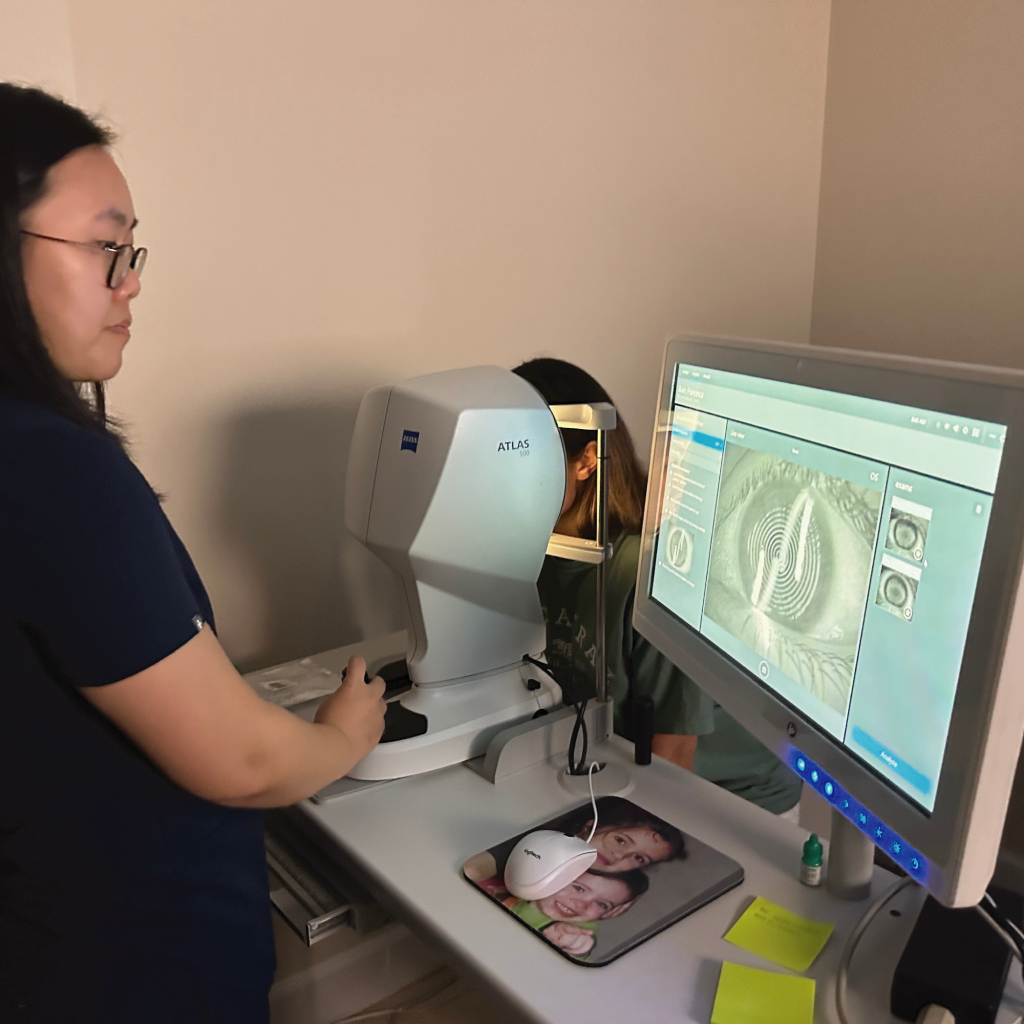
Computerized corneal topography is a noncontact imaging test that creates color-coded maps of the curvature and shape of the cornea, the clear front surface of the eye. The maps help an eye-care professional evaluate regular and irregular astigmatism, compare corneal shape over time, and plan selected forms of vision correction.
Topography is one part of an eye examination. A map does not establish a diagnosis, prove that a condition is progressing, or determine the final contact lens design by itself. Dr. Edward Boshnick interprets the measurements together with the patient’s symptoms, refraction, eye-health findings, prior procedures, and other imaging when needed.
What Happens During Corneal Topography?
The patient rests the chin and forehead against the instrument and looks at a fixation target. A Placido-based topographer projects a pattern of rings onto the tear film covering the cornea and analyzes the reflected pattern. The instrument converts those measurements into maps that can be reviewed on a computer.
No instrument touches the eye during routine Placido-based topography. Several captures may be taken because blinking, tear-film instability, fixation, eyelid position, and movement can affect map quality. The clinician checks whether a scan is reliable before comparing or interpreting it.
Contact lenses can temporarily alter corneal shape. Follow the practice’s instructions about whether and when to remove current lenses before testing; the appropriate interval depends on the lens type, reason for the scan, cornea, and clinical question. Bring prior scans and lens information when available.
What the Color-Coded Maps Can Show
Corneal topography can show areas that are relatively steep, flat, regular, asymmetric, or irregular. Depending on the map type and scale, warmer colors often represent steeper curvature and cooler colors flatter curvature. The colors must be interpreted with the numerical values, map scale, scan quality, and the rest of the examination; color alone is not a diagnosis.
Topography may contribute to the evaluation of keratoconus, corneal ectasia, irregular astigmatism, corneal changes after LASIK or RK, and the optical effect of scarring or transplantation. Reliable maps obtained over time may help identify change, but apparent differences can also result from tear-film quality, contact lens warpage, or inconsistent capture.
Corneal Topography vs. Corneal Tomography
These terms are related but not interchangeable. Placido-based topography primarily measures curvature of the front corneal surface. Corneal tomography can evaluate the front and back corneal surfaces and provide corneal-thickness information. Dr. Boshnick selects and combines tests according to the condition and question being evaluated.
How Topography Supports Custom Scleral Lens Fitting
For a person being evaluated for custom scleral lenses, topography helps describe the corneal irregularity that the fluid-filled lens is intended to vault. It can help Dr. Boshnick understand cone location, asymmetry, post-surgical shape, and how much blur may arise from the front corneal surface.
Corneal topography does not map the complete scleral landing area and cannot confirm a final scleral lens fit on its own. A diagnostic lens must be evaluated on the eye after it settles. Clearance, limbal relationship, scleral landing, centration, rotation, surface quality, vision, comfort, and eye health all remain important. Anterior-segment OCT or other measurements may provide additional information for selected eyes.
The scleral lens fitting guide explains how mapping, a diagnostic lens trial, refraction, handling instruction, and follow-up work together.
How Dr. Boshnick Uses Corneal Mapping
Dr. Boshnick uses corneal mapping to answer a defined clinical question rather than treating the map as a stand-alone result. Depending on the patient, he may compare topography with refraction, slit-lamp examination, tear-film and ocular-surface findings, prior surgical records, tomography, anterior-segment OCT, wavefront measurements, and an on-eye diagnostic lens assessment.
Not every patient requires every technology. Topography does not guarantee scleral lens candidacy, predict a particular visual result, or replace medical monitoring or treatment of an active corneal condition. When progression, corneal cross-linking, or surgery is a concern, care may also involve a corneal ophthalmologist.
Request an evaluation with Dr. Boshnick or call 305-271-8206. Patients traveling from outside the area should contact the practice before making arrangements because measurements and diagnostic lens testing require an in-person examination.
It is a noncontact imaging test that creates color-coded maps of the curvature and shape of the front corneal surface. The maps are interpreted with the examination and other measurements.
It may be used to evaluate irregular astigmatism, keratoconus or ectasia, changes after corneal surgery, contact lens effects, or corneal shape relevant to specialty lens planning and monitoring.
You rest your chin and forehead at the instrument and look at a target while it captures reflected light patterns from the corneal surface. Several scans may be taken to confirm measurement quality.
Follow the practice’s specific instructions. Contact lenses can temporarily alter corneal shape, but the appropriate removal interval depends on the lens type, cornea, and reason for testing.
Placido-based topography primarily maps curvature of the front corneal surface. Tomography can evaluate the front and back surfaces and provide thickness information. The tests may complement each other.
It describes corneal shape and irregularity that a scleral lens must vault. It does not map the complete scleral landing area or replace an on-eye diagnostic lens assessment after settling.
No. Diagnosis and progression assessment use the history, examination, reliable imaging, thickness and posterior-surface information when indicated, and comparison over time. Tear-film instability or contact lens warpage can affect a map.