7800 SW 87 AVE SUITE B-270 MIAMI, FLORIDA 33173


- Home
- Radial Keratotomy Complications
Radial Keratotomy Complications
Understanding Radial Keratotomy Complications
Radial keratotomy (RK) was an older refractive surgery that used spoke-like corneal incisions to reduce nearsightedness. Those incisions permanently changed the cornea. If vision becomes blurry, distorted, or less predictable years later, possible contributors include a shift toward farsightedness, irregular astigmatism, tear-film or ocular-surface disease, cataract, or another eye condition. A symptom alone cannot identify the cause.
Long-term change is well documented. In the National Eye Institute-supported 10-year PERK study, more than 40% of RK-operated eyes continued to shift toward farsightedness. Studies have also documented changes in refraction and corneal shape across the day. Patients may describe ghosting, multiple images, glare, halos, starbursts, reduced contrast, or a prescription that does not remain consistent.
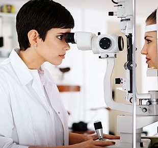
Dr. Edward Boshnick evaluates the cornea, ocular surface, current prescription, and overall eye health before recommending a visual rehabilitation option. When irregular corneal optics are an important part of the problem and glasses or standard soft contacts do not provide useful vision, a custom scleral lens may be considered. A scleral lens can create a new front optical surface, but it does not repair or reverse RK.
Common reasons patients seek an evaluation after RK include:
- Progressive hyperopic shift: The eye may become more farsighted over time, changing distance and near prescriptions.
- Fluctuating or blurry vision: Clarity may change from morning to evening, from one day to another, or with ocular-surface conditions.
- Irregular astigmatism: An uneven corneal shape can cause distortion that glasses or standard soft lenses may not fully correct.
- Glare, halos, and starbursts: Light scatter and higher-order optical aberrations may be more noticeable at night or in dim conditions.
- Ghosting or multiple images: Irregular corneal optics can create overlapping or shadowed images, sometimes in one eye.
- Dry-eye or ocular-surface symptoms: Burning, irritation, light sensitivity, or unstable vision may coexist with the post-RK corneal shape.
- Difficulty obtaining a stable correction: A glasses or contact-lens prescription may not capture the full range of daily visual change.
- Cataract or another eye condition: Not every new symptom comes from the RK cornea, especially decades after the original surgery.
- Corneal-health concerns: Old incision sites remain structurally different, so trauma, sudden pain, redness, or abrupt vision loss needs prompt evaluation.
Why Can Vision Change Years After RK?
RK flattened the central cornea by changing its biomechanics with deep radial incisions. The result may continue to evolve over time. A gradual hyperopic shift means the eye moves toward farsightedness. Diurnal fluctuation means refraction or corneal shape changes across the day. The direction and amount of fluctuation are not identical for every patient, which is why a single glasses measurement may not explain the full experience.
Irregular astigmatism can also scatter light and create blur, ghosting, glare, halos, or reduced contrast. Dry eye can make the optical surface less stable. Cataract, retinal disease, and other age-related changes may produce similar complaints and must be considered rather than assuming every symptom is an RK complication.
How Is Blurry Vision After RK Evaluated?
A useful evaluation begins by identifying which part of the eye is limiting vision and whether the prescription changes meaningfully over time. Depending on the history and findings, the assessment may include:
- Visual acuity and refraction: measurements with glasses and, when useful, at different times of day.
- Slit-lamp examination: assessment of the incision pattern, corneal clarity, ocular surface, tear film, and other front-of-eye structures.
- Corneal topography or tomography: maps that help show central flattening, steep and flat regions, asymmetry, and irregular astigmatism. Learn how computerized corneal topography supports specialty-lens planning.
- Pachymetry: measurement of corneal thickness, which can be important when monitoring a post-RK cornea and its response to lens wear.
- Ocular-surface assessment: review of dryness, eyelids, tear quality, staining, and prior treatment.
- Lens, retina, and optic-nerve assessment: evaluation for cataract or another condition that a contact lens cannot correct.
- Diagnostic specialty-lens trial: an on-eye trial may help show how much of the visual limitation comes from irregular corneal optics.
What Options Are Available After Radial Keratotomy?
There is no single option that is right for every post-RK eye. The plan depends on the source of blur, the degree of fluctuation, corneal and ocular-surface health, cataract status, visual goals, and the response to diagnostic correction.
- Glasses may help when the prescription is sufficiently regular and stable.
- Soft, corneal gas-permeable, hybrid, or scleral lenses may be considered based on corneal shape, comfort, stability, handling ability, and eye-health findings.
- Dry-eye or ocular-surface care may be needed when tear-film disease contributes to blur or discomfort. See the dry-eye evaluation guide.
- Corneal or cataract ophthalmology evaluation may be appropriate when structural disease, cataract, or a surgical question is the main concern. Prior RK can make cataract measurements and refractive expectations more complex.
The phrase “RK repair” can mean different things. No lens removes the incisions or returns the cornea to its pre-RK structure. A careful examination is needed before discussing additional surgery because treating a prescription, an irregular cornea, a cataract, and an ocular-surface problem are different tasks.
How Can Scleral Lenses Help After RK?
A scleral lens is a large-diameter, oxygen-permeable rigid lens that rests on the sclera and vaults over the cornea. It is filled with sterile, preservative-free saline before insertion. The rigid front surface and fluid reservoir can mask part of the irregular corneal optics, which may reduce blur, ghosting, or distortion for selected patients.
Because the lens lands beyond the cornea, it can avoid direct central bearing on the RK incision pattern. This does not mean the lens is automatically appropriate or that every post-RK eye will obtain the same vision, comfort, or wearing time. Learn more in the focused guide to scleral lenses after LASIK and RK.
Why Post-RK Scleral Lens Fitting Needs Extra Care
Post-RK corneas are often oblate, with a flatter center and relatively steeper midperiphery, and the shape may be asymmetric. Lens diameter, sagittal depth, central and limbal clearance, scleral landing, settling, optics, and tear-reservoir depth therefore need patient-specific assessment.
A published case series of nine post-RK eyes found measurable scleral-lens-induced corneal edema, with greater magnitude and variability toward the midperiphery near the approximate incision locations. This small study does not mean scleral lenses are unsafe for every post-RK patient. It does support careful baseline measurement, oxygen-conscious design, and assessment of the cornea after the lens has been worn.
Follow-up may include review of:
- central and limbal clearance after the lens settles;
- landing-zone alignment, edge compression, and lens stability;
- lens material, lens thickness, fluid-reservoir depth, and other factors that affect oxygen delivery;
- bubbles, debris, midday fogging, surface wetting, comfort, and visual stability;
- corneal thickness, clarity, staining, redness, and other eye-health findings after wear; and
- safe insertion and removal without unnecessary force or suction.
The scleral lens fitting guide explains what patients can expect during diagnostic trials and follow-up.
What Scleral Lenses Cannot Do
- They do not close RK incisions or restore the original corneal structure.
- They do not reverse hyperopic shift or guarantee that the eye will stop changing.
- They do not guarantee normal or perfectly stable vision throughout the day.
- They do not treat cataract, retinal disease, active infection, or another non-corneal cause of vision loss.
- They do not replace diagnosis-specific medical care or follow-up.
Dr. Edward Boshnick’s Post-RK Evaluation
Dr. Boshnick’s role is to determine whether specialty optics can safely address the corneal component of a patient’s visual problem. He reviews the surgical and contact-lens history, current refraction, corneal maps, ocular-surface findings, diagnostic lens response, and the lens-eye relationship after settling. When a conventional scleral lens provides a stable platform but higher-order aberrations remain, wavefront-guided scleral optics may be evaluated for selected eyes.
Some patients need coordinated care with a corneal, cataract, retinal, or other eye-care specialist. To discuss whether a post-RK specialty-lens evaluation is appropriate, contact Eye Freedom. Patients with broader concerns can also review post-surgical vision loss and rehabilitation.
When Should a Post-RK Patient Seek Prompt Care?
Sudden vision loss, new or increasing eye pain, pronounced redness, discharge, marked light sensitivity, a new white or cloudy area on the cornea, or symptoms after eye trauma should not wait for a routine contact-lens visit. Remove a lens if it is being worn and seek prompt professional eye care.
Medical sources
- National Eye Institute: 10-year results of the PERK study
- NCBI Bookshelf: Radial Keratotomy Correction
- PubMed: Scleral lens-induced corneal edema after radial keratotomy
- Peer-reviewed review of radial keratotomy and current management
- Peer-reviewed review of scleral lenses, indications, and complications
This page is educational and does not provide a diagnosis or individualized treatment plan. Recommendations depend on an examination of the individual eye.
Your Vision, Our Priority:
Schedule Your Consultation Now
Frequently Asked Questions
Radial keratotomy, or RK, is an older refractive procedure that used spoke-like, partial-thickness corneal incisions to flatten the central cornea and reduce nearsightedness. The incisions permanently changed the corneal structure and biomechanics.
Possible causes include progressive hyperopic shift, irregular astigmatism, daily corneal-shape fluctuation, dry eye, cataract, retinal disease, or another eye condition. An examination is needed because the symptom does not identify the cause by itself.
Studies have documented diurnal changes in refraction and corneal topography after RK. The amount and direction can vary among patients, and tear-film changes may add further fluctuation. Measurements at more than one time may be useful when symptoms and a single refraction do not match.
RK cannot simply be reversed because the incisions permanently changed the cornea. Glasses, specialty lenses, ocular-surface care, cataract care, or selected corneal procedures may address a particular problem, but none returns the cornea to its original pre-RK structure.
Options may include glasses, soft lenses, corneal gas-permeable lenses, hybrid lenses, scleral lenses, ocular-surface treatment, or referral for a cataract or corneal evaluation. The appropriate choice depends on the cause of blur, corneal health, prescription stability, visual goals, and diagnostic testing.
For selected patients, a scleral lens can vault the incision-bearing cornea and create a regular front optical surface that may reduce blur, ghosting, or distortion. It does not close the incisions, reverse RK, stop future corneal change, or guarantee a particular result.
They may be appropriate for selected eyes, but safety cannot be assumed. Post-RK fitting requires assessment of corneal health, clearance, settling, landing, oxygen-related factors, and the eye after lens wear. Published evidence of regional edema near RK incisions supports individualized monitoring.
The evaluation may include visual acuity, refraction, slit-lamp examination, corneal topography or tomography, pachymetry, ocular-surface assessment, and a diagnostic lens trial. Cataract, retinal, and optic-nerve findings are also considered because a scleral lens cannot correct every cause of reduced vision.
No. Some patients obtain useful vision with glasses, soft lenses, corneal gas-permeable lenses, or hybrid lenses. Others need treatment for dry eye, cataract, or another condition. A diagnostic assessment helps determine whether a scleral lens provides a worthwhile and medically acceptable option.
Seek prompt professional care for sudden vision loss, new or increasing pain, pronounced redness, discharge, marked light sensitivity, a new white or cloudy corneal area, or symptoms after eye trauma. These concerns should not wait for a routine lens visit.





Schedule Your Consultation with
Dr. Boshnick
-
-
-
7800 Sw 87 Ave Suite B-270
Miami, Florida 33173
Schedule Your Consultation with
Dr. Boshnick
-
-
-
7800 Sw 87 Ave Suite B-270
Miami, Florida 33173
