7800 SW 87 AVE SUITE B-270 MIAMI, FLORIDA 33173


- Home
- Cirrus Optical Coherence Tomographer (OCT)
Cirrus Optical Coherence Tomographer (OCT)
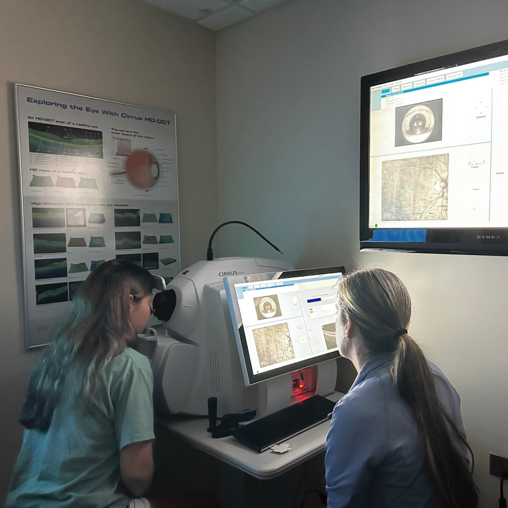
Cirrus optical coherence tomography (OCT) is a noncontact eye-imaging test that uses reflected light to create cross-sectional maps of the retina and optic nerve. At Eye Freedom, Dr. Edward Boshnick may use ZEISS Cirrus OCT when the macula, retinal layers, retinal nerve fiber layer, ganglion cells, or optic nerve are relevant to an evaluation.
The scan adds structural information to the patient’s history and eye examination. It does not treat disease, establish every diagnosis, prove progression, or determine a treatment plan by itself.
What Does a Cirrus OCT Eye Scan Measure?
OCT measures how light reflects from tissue at different depths. Software converts that information into cross-sectional images, thickness maps, and three-dimensional data sets. The selected scan protocol determines which structures and measurements are displayed.
- Macular scans can show the contour and measured layers of the central retina. Depending on the clinical question, they may help document swelling, traction, thinning, or another structural feature that needs interpretation.
- Optic-nerve scans can measure the retinal nerve fiber layer (RNFL) and selected optic-nerve-head features.
- Ganglion-cell analysis can measure selected inner retinal layers around the macula that may add context to an optic-nerve or glaucoma evaluation.
On an order or report, OCT macula Cirrus OU generally means that a Cirrus macular OCT protocol was performed for both eyes; OU is the chart abbreviation for both eyes. That wording identifies the test and laterality, not the result or a diagnosis.
Some reports compare measurements with a reference database and display color-coded ranges. A color flag is a statistical comparison, not proof of disease. Age, anatomy, refractive error, image quality, and the limits of the reference population can all affect interpretation.
What Happens During a Cirrus OCT Scan?
The imaging usually takes only a few minutes. You sit at the instrument with your chin and forehead supported while one eye is imaged at a time. The operator aligns the scan and asks you to look toward a fixation target. You may be asked to blink before a capture, hold still briefly, or repeat a scan if movement, blinking, or poor alignment reduces image quality.
The scanner normally does not touch the eye and uses light rather than ionizing radiation. The target or scanning light may appear bright, but the imaging itself should not require an incision or injection.
Will My Eyes Need To Be Dilated?
Not every OCT scan requires dilation. Dr. Boshnick may recommend dilating drops when pupil size, cataract or another media opacity, the clinical question, or the rest of the internal-eye examination makes a wider pupil useful. If dilation is used, near vision may be temporarily blurred and the eyes may be light-sensitive for several hours. Follow the practice’s driving instructions for your visit.
Cirrus OCT vs. Retinal Photography
Color fundus photography records the visible surface appearance of the retina, optic nerve, and retinal blood vessels. OCT creates cross-sectional images and measurements of selected retinal or optic-nerve layers. The tests are complementary rather than interchangeable, and one or both may be selected depending on the clinical question.
Why Serial OCT Comparisons Need Clinical Review
Repeat scans can help document whether a measured feature appears stable or has changed. Apparent change can also result from a different scan position, blink or eye-movement artifact, poor fixation, a dry ocular surface, cataract or another opacity, low signal strength, anatomy outside the reference database, or an automated segmentation error.
For that reason, an OCT change map should not be treated as automatic proof of progression. Dr. Boshnick reviews image quality and the underlying cross-sectional scans, then compares the findings with symptoms, visual acuity, eye pressure when indicated, the dilated examination, photographs, and visual field testing. Additional testing or referral may be necessary.
How Dr. Boshnick Uses Cirrus OCT
Dr. Boshnick selects imaging according to the patient’s history, symptoms, previous diagnoses, medications, prior surgery, examination findings, and the question that needs to be answered. A baseline OCT may be useful in selected cases, while another patient may need a different test or no OCT at that visit.
When a scan or examination raises concern about retinal disease, glaucoma, optic-nerve disease, or another condition requiring medical or surgical care, Dr. Boshnick may recommend referral or co-management with an appropriate ophthalmologist or other specialist. OCT supports that decision; it does not replace the specialist evaluation.
Cirrus OCT on this page primarily addresses retinal and optic-nerve imaging. For corneal anatomy, the front of the eye, and selected scleral-lens relationships, see Eye Freedom’s guide to Visante anterior-segment OCT.
When Not To Wait for a Routine Scan
Sudden vision loss, a new curtain or shadow, a sudden shower of floaters or flashes, severe eye pain, significant eye injury, or a red painful eye needs urgent in-person assessment. Do not wait for a routine OCT appointment or remote image review when these symptoms occur.
Request an evaluation with Dr. Boshnick or call 305-271-8206 to discuss the appropriate next step. Patients traveling from another state or country should contact the practice before making arrangements because imaging and examination needs vary.
Medical and Device Sources
- National Eye Institute: Glaucoma detection and comprehensive eye examination
- ZEISS: CIRRUS OCT platform and imaging capabilities
- Peer-reviewed study: OCT artifacts and interpretation of glaucoma progression
- Peer-reviewed review: Avoiding OCT misinterpretation and artifacts
This page is educational. It does not interpret an individual scan, diagnose a condition, or recommend treatment.
Your Vision, Our Priority:
Schedule Your Consultation Now
Frequently Asked Questions
A Cirrus OCT eye scan is a noncontact imaging test that uses reflected light to create cross-sectional images and measurements of selected retinal or optic-nerve structures. The results are interpreted with the patient’s history and eye examination.
It generally means that a Cirrus OCT macular scan was performed for both eyes. OU is the chart abbreviation for both eyes. The phrase identifies the test and laterality; it does not state whether the result is normal or provide a diagnosis.
Depending on the selected protocol, it can measure the retinal nerve fiber layer and selected optic-nerve-head or ganglion-cell features. These measurements can add structural information, but they must be interpreted with the examination and other testing.
The scanner normally does not touch the eye and uses light rather than ionizing radiation. You rest your chin and forehead at the instrument and look toward a target while one eye is imaged at a time.
Not always. Dilation may be recommended because of pupil size, image quality, cataract or another opacity, the clinical question, or the rest of the internal-eye examination. Dilating drops can temporarily blur near vision and increase light sensitivity.
A retinal photograph records the visible surface appearance of the retina, optic nerve, and blood vessels. OCT creates cross-sectional images and measurements of selected tissue layers. The tests answer different questions and may be used together.
No. OCT can provide useful structural measurements, but glaucoma assessment also considers the history, optic-nerve examination, eye pressure, visual fields, scan quality, and other findings. Apparent OCT change must be checked for artifacts and clinical agreement.
Yes. Blinking, eye movement, poor fixation, a dry ocular surface, cataract or another opacity, low signal, scan position, and automated segmentation errors can affect image quality or measurements. The clinician should review the underlying scans rather than relying only on color flags or automated summaries.
Schedule Your Consultation with
Dr. Boshnick
-
-
-
7800 Sw 87 Ave Suite B-270
Miami, Florida 33173
Schedule Your Consultation with
Dr. Boshnick
-
-
-
7800 Sw 87 Ave Suite B-270
Miami, Florida 33173
