7800 SW 87 AVE SUITE B-270 MIAMI, FLORIDA 33173


- Home
- Advanced Digital Retinal Imaging: A Patient Guide
Advanced Digital Retinal Imaging: A Patient Guide
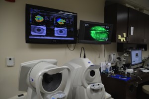
Advanced digital retinal imaging creates detailed pictures of the retina, macula, optic nerve, and retinal blood vessels at the back of the eye. The term describes a group of tests rather than one single scan. Each method answers a different clinical question, and the images are interpreted together with the patient’s history and eye examination.
Dr. Edward Boshnick uses retinal images to document relevant findings, establish a baseline when appropriate, compare changes over time, and decide whether additional testing or referral is needed. Imaging adds information; it does not by itself provide every diagnosis or replace every part of a comprehensive eye examination.
What Is Advanced Digital Retinal Imaging?
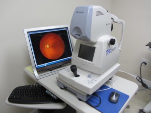
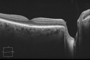
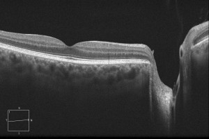
Color fundus photography uses a specialized camera to photograph the visible back of the eye. It can document the retina, macula, optic nerve, and retinal blood vessels. Optical coherence tomography (OCT) uses reflected light to create cross-sectional images of retinal or optic-nerve tissue. A fundus photograph and an OCT scan are not interchangeable: one records surface appearance and the other shows measured layers beneath that surface. Learn more about Eye Freedom’s Cirrus optical coherence tomography.
For either test, the patient usually sits at the instrument, rests the forehead and chin, and looks toward a target while images are captured. A camera may produce a bright flash, while OCT uses a scanning light. These methods do not use ionizing radiation and usually do not touch the eye. Pupil dilation may still be recommended depending on the clinical question, pupil size, image quality, and the view needed for the examination. Dilating drops can temporarily blur near vision and increase light sensitivity.
What Can Retinal Images Document?
Retinal imaging can help document the appearance of the retina, central macula, optic nerve, and blood vessels. Depending on the type and quality of the image, it may show findings associated with diabetic retinopathy, age-related macular degeneration, retinal vascular disease, inflammation, or an optic-nerve appearance that warrants a glaucoma evaluation.
An image is one part of the evidence. It may not capture the entire peripheral retina, and cataract, a small pupil, eye movement, poor fixation, or other factors can reduce image quality. A normal-looking photograph cannot rule out every eye disease. Findings may need confirmation with dilation, OCT, pressure measurement, visual field testing, laboratory testing, or evaluation by an ophthalmologist or retina specialist.
Why Compare Retinal Images Over Time?
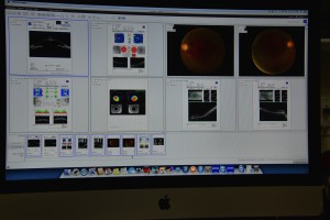
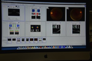
A baseline image provides a dated record that can be compared with later photographs or scans. Serial comparison may help an eye doctor recognize whether a documented feature appears stable, new, or changed. This can be useful when following selected retinal, macular, or optic-nerve findings.
Small apparent differences do not always represent disease progression. Scan position, field of view, pupil size, focus, segmentation, instrument type, and image quality can affect the comparison. Dr. Boshnick interprets serial images in the context of symptoms, examination findings, and other measurements. The appropriate interval is individualized rather than automatically annual for every patient.
How Telemedicine Can Support Image Review
When clinically appropriate, securely transmitted retinal images and records may support pre-visit planning, care coordination, follow-up decisions, or referral triage. This is often called store-and-forward telemedicine: an image is captured first and reviewed later with the relevant history and records.
Remote image review is not a substitute for every in-person examination. Its usefulness depends on the reason for review, image type, image quality, field of view, supporting information, patient location, consent, privacy safeguards, and whether hands-on testing is needed. A patient-supplied photograph or an isolated scan may be insufficient for diagnosis or treatment decisions.
Patients traveling from another state or country may send available records before arranging an evaluation, but remote review cannot guarantee candidacy, eliminate required testing, or promise that one visit will answer every clinical question. Sudden vision loss, a new curtain or shadow, a sudden shower of floaters or flashes, severe eye pain, significant injury, or a red painful eye requires urgent in-person care rather than a routine telemedicine request.
How Dr. Boshnick Uses Retinal Imaging
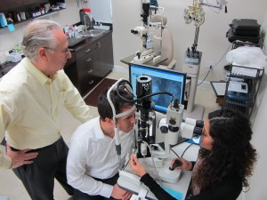
Dr. Boshnick reviews retinal images alongside the patient’s history, visual acuity, pupils, eye pressure when indicated, slit-lamp examination, and dilated internal-eye examination when needed. OCT, visual field testing, or another study may be added when it answers a specific question. If an image or examination suggests a retinal, neurologic, glaucoma, or other medical concern outside the appropriate scope of the visit, he may recommend referral or co-management.
Before an evaluation, bring current glasses and contact lenses, a medication list, prior diagnoses, retinal photographs, OCT or visual-field reports, and relevant surgical or specialist records when available. Contact the practice before traveling so the team can explain which records may be useful.
Request an evaluation with Dr. Boshnick or call 305-271-8206. The original consultation form appears below.
Medical Sources
- National Eye Institute: Dilated eye examination
- American Society of Retina Specialists: Advanced retinal imaging patient guide
- American Optometric Association: 2025 eye and vision telehealth position statement
- Systematic review: Digital retinal photography for diabetic-retinopathy screening
- Systematic review and meta-analysis: Teleophthalmology diagnostic performance
This page is educational and does not diagnose a condition, interpret an individual image, or replace an examination. The appropriate tests and follow-up depend on the patient’s history and findings.
Your Vision, Our Priority:
Schedule Your Consultation Now
Frequently Asked Questions
It is an umbrella term for technologies that create digital images of structures at the back of the eye, including the retina, macula, optic nerve, and retinal blood vessels. Different tests provide different information and must be interpreted with the examination.
No. Fundus photography captures a color image of the visible back of the eye. OCT uses reflected light to create cross-sectional images of retinal or optic-nerve layers. The tests answer different questions and may complement one another.
No. Retinal images can add valuable documentation, but they do not automatically show the entire peripheral retina or replace the other parts of a comprehensive examination. Dr. Boshnick determines whether dilation and additional testing are needed.
Not always. Some photographs and OCT scans can be captured without drops, but dilation may be needed because of pupil size, image quality, the clinical question, or the view required for the examination.
Depending on the image type, retinal imaging may document findings associated with diabetic retinopathy, macular degeneration, retinal vascular disease, inflammation, or optic-nerve and glaucoma concerns. Imaging alone does not establish every diagnosis.
There is no single interval for everyone. Timing depends on age, symptoms, medical and family history, previous findings, risk factors, and the condition being followed. Dr. Boshnick recommends testing based on the individual examination.
Selected images may support secure pre-visit review, care coordination, follow-up planning, or referral triage. Usefulness depends on image quality, supporting records, the reason for review, and whether an in-person examination is required.
Seek urgent in-person care for sudden vision loss, a new curtain or shadow, a sudden shower of floaters or flashes, severe eye pain, significant eye injury, or a red painful eye. Do not wait for routine telemedicine review of these symptoms.
Schedule Your Consultation with
Dr. Boshnick
-
-
-
7800 Sw 87 Ave Suite B-270
Miami, Florida 33173
Schedule Your Consultation with
Dr. Boshnick
-
-
-
7800 Sw 87 Ave Suite B-270
Miami, Florida 33173
