7800 SW 87 AVE SUITE B-270 MIAMI, FLORIDA 33173


- Home
- Corneal Ectasia
Corneal Ectasia
What Is Corneal Ectasia?
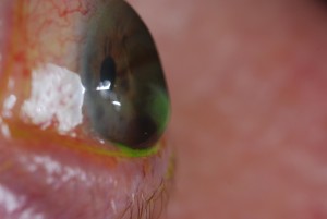
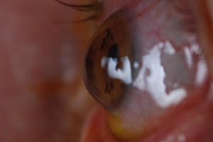
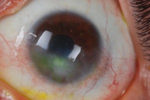
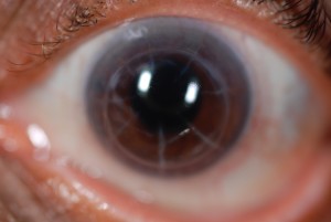
Corneal ectasia is a group of conditions in which the cornea becomes thinner, steeper, or more irregular. Because the cornea is the eye’s main front focusing surface, these changes can create irregular astigmatism and vision that is blurred, distorted, ghosted, or difficult to correct with glasses.
Ectasia can be associated with keratoconus, pellucid marginal degeneration, or changes after refractive surgery such as LASIK or PRK. Post-refractive ectasia is uncommon, but it may develop months or years after surgery. The diagnosis and rate of change differ from person to person.
How Corneal Ectasia Affects Vision
An ectatic cornea does not bend light evenly. As its shape becomes more irregular, a standard glasses prescription may correct only part of the optical problem. Some patients notice increasing nearsightedness or astigmatism, while others report glare, halos, starbursts, shadow images, or reduced contrast, especially at night.
These symptoms are not specific to ectasia and can also occur with dry eye, cataract, retinal disease, or other conditions. A comprehensive examination and corneal imaging are needed to identify the cause. Patients with worsening vision after LASIK or PRK may also find the LASIK complications guide helpful.
Symptoms and Causes
Early ectatic change may be subtle, and symptoms do not always match the amount of change seen on a scan. Findings that may lead an eye-care professional to evaluate the cornea more closely include:
- Increasing nearsightedness or astigmatism: A prescription that changes repeatedly can be a sign that the corneal shape is changing.
- Blurred, fluctuating, or distorted vision: Vision may vary by eye, lighting, time of day, or method of correction.
- Ghosting or multiple images: A person may see a faint shadow or duplicate edge around letters, lights, or objects.
- Glare, halos, and starbursts: These symptoms may be more noticeable around headlights or other bright lights at night.
- Reduced vision with glasses or soft lenses: Conventional correction may become less effective as irregular astigmatism increases.
- Contact lens instability or intolerance: A changing or irregular corneal surface can make some conventional lenses less stable or comfortable.
- Relevant history or risk factors: Keratoconus, eye rubbing, allergic eye disease, family history, and prior refractive surgery may be clinically relevant, but none confirms ectasia by itself.
How Corneal Ectasia Is Diagnosed
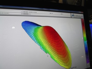
Diagnosis is based on the history, refraction, slit-lamp examination, and measurements of corneal shape and thickness. Corneal topography maps the front surface of the cornea, while tomography can evaluate the front and back surfaces and provide pachymetry data. Comparing reliable scans over time helps determine whether the condition appears stable or progressive.
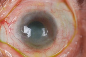
Dr. Edward Boshnick evaluates the optical effect of an irregular cornea and whether a specialty lens may improve functional vision. When progression, cross-linking, or surgery is a concern, care may also involve a corneal ophthalmologist. Worsening vision, new pain, marked light sensitivity, or sudden corneal clouding should be evaluated promptly.
Treatment, Monitoring, and Scleral Lenses
Management depends on the cause, severity, documented progression, corneal thickness, age, ocular surface, and visual needs. Glasses or conventional contact lenses may be adequate in milder cases. Corneal cross-linking may be considered by a corneal specialist to slow or stabilize progressive ectasia, but it does not guarantee that the cornea will regain a regular shape or that specialty correction will no longer be needed. Other procedures may be considered in selected cases.
How Scleral Lenses May Help
A scleral lens is a large-diameter gas-permeable lens that vaults the cornea and rests on tissue over the sclera. Its fluid-filled space and front optical surface can mask some corneal irregularity and may provide clearer, more stable vision for selected patients. A scleral lens does not reverse ectasia or stop progression, so continued corneal monitoring remains important.
Lens candidacy depends on corneal and ocular surface health, clearance, landing, oxygen considerations, handling ability, comfort, and the visual response during an on-eye evaluation. Explore Eye Freedom’s guides to scleral lenses for keratoconus and scleral lenses for ectasia and irregular corneas. In optically complex cases, Dr. Boshnick may also assess whether wavefront-guided scleral lenses are appropriate.
Request an evaluation with Eye Freedom.
Medical Sources
- Peer-reviewed systematic review: corneal ectasia after refractive surgery
- Peer-reviewed review: risk factors for ectasia after corneal refractive surgery
- Peer-reviewed meta-analysis: corneal cross-linking for post-refractive ectasia
- American Academy of Ophthalmology report: cross-linking for progressive ectasia
- Peer-reviewed review: scleral lenses in corneal ectasia
This page is educational and does not provide a diagnosis or individualized treatment plan. An examination is needed to determine the cause of symptoms, whether the cornea is changing, and which options may be appropriate.
Your Vision, Our Priority:
Schedule Your Consultation Now
Frequently Asked Questions
Corneal ectasia describes thinning, steepening, or irregular reshaping of the cornea. The change can produce irregular astigmatism and blurred, ghosted, or distorted vision that glasses may not fully correct.
Keratoconus is one form of corneal ectasia. Ectatic change can also occur with pellucid marginal degeneration or after refractive surgery such as LASIK or PRK. The conditions share some optical effects but may have different histories and management considerations.
An eye-care professional considers symptoms, prescription changes, slit-lamp findings, corneal topography or tomography, and corneal-thickness measurements. Reliable scans compared over time can help determine whether the condition appears stable or progressive.
Corneal cross-linking may slow or stabilize progression in appropriately selected patients, but individual results vary. It is not intended to restore a perfectly regular corneal shape, and a corneal specialist must determine whether it is appropriate.
No. Scleral lenses are used for optical correction and, in selected cases, ocular surface support. They do not reverse ectasia or stop progression, so corneal monitoring and any recommended medical or surgical care remain important.
A properly fitted scleral lens vaults the irregular cornea and provides a new front optical surface. For selected patients, this may improve visual clarity and stability when glasses or conventional contact lenses do not provide adequate correction.
Arrange an examination when blur, ghosting, glare, prescription changes, or reduced corrected vision is new or worsening. New pain, marked light sensitivity, or sudden corneal clouding warrants prompt professional evaluation.
Schedule Your Consultation with
Dr. Boshnick
-
-
-
7800 Sw 87 Ave Suite B-270
Miami, Florida 33173
Schedule Your Consultation with
Dr. Boshnick
-
-
-
7800 Sw 87 Ave Suite B-270
Miami, Florida 33173
