7800 SW 87 AVE SUITE B-270 MIAMI, FLORIDA 33173

Contact Us
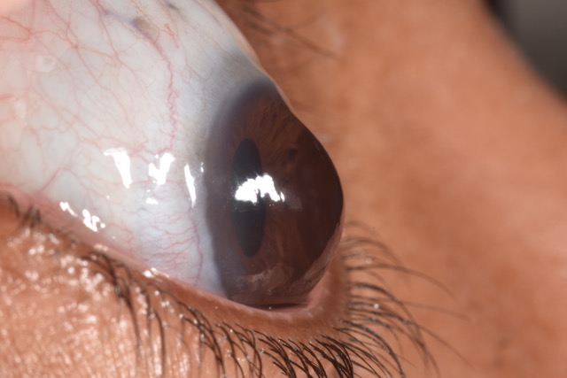
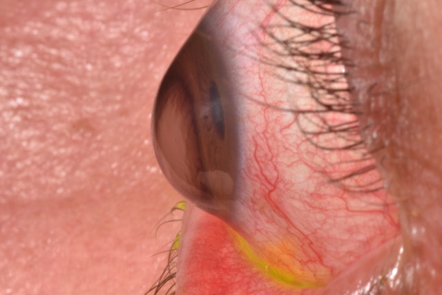
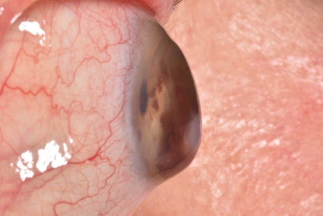
Keratoconus is a condition in which the cornea becomes thinner and develops an irregular, cone-like shape. Because the cornea provides much of the eye’s focusing power, this change can produce blur, ghosting, glare, irregular astigmatism, and vision that glasses do not fully correct.
Keratoconus often begins during the teenage years or young adulthood, although the age of diagnosis and rate of change vary. It usually affects both eyes unevenly. It is not contagious, and having keratoconus does not automatically mean that a person will need surgery or lose functional vision.
Care has two separate goals: monitor the cornea and address progression, and provide the clearest, most comfortable vision possible. The right plan depends on corneal measurements, change over time, ocular surface health, symptoms, and prior correction.
Keratoconus can look different from one person to another. Symptoms may include:
These symptoms are not specific to keratoconus, so diagnosis requires an eye examination and corneal measurements. Our keratoconus signs guide explains when a specialty evaluation may be useful.
Seek prompt eye care for a sudden drop in vision, new pain, pronounced redness, marked light sensitivity, or sudden corneal clouding.
Keratoconus is considered multifactorial, which means there is usually no single identifiable cause. Genetics can contribute, and the condition is more common in some families. Eye rubbing and allergic or atopic disease are also associated with keratoconus and may be relevant when an eye-care professional evaluates risk and progression.
A person did not cause keratoconus by reading, using screens, or wearing glasses. Patients with itching or habitual eye rubbing should discuss those symptoms with their eye-care team rather than trying to manage them without guidance.
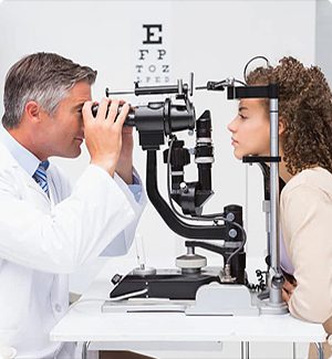
An evaluation may include refraction, visual-acuity testing, slit-lamp examination, and a review of prescription changes and family history. Corneal topography maps the curvature of the front surface. Tomography can evaluate the front and back of the cornea and its thickness distribution. These measurements may identify changes that are not obvious during a routine vision screening.
One examination shows the eye’s current shape; comparison over time helps determine whether the condition is stable or progressing. Monitoring intervals should be individualized according to age, measurements, symptoms, and prior change.
Improving vision and slowing progression are not the same job.
Read the non-surgical keratoconus treatment guide for a fuller comparison of these options.
A scleral lens is a large-diameter, oxygen-permeable rigid lens. It rests on the sclera, the white part of the eye, and vaults over the irregular cornea. The space between the lens and cornea is filled with preservative-free saline before insertion.
This design creates a new, regular optical surface that may reduce blur and distortion caused by irregular corneal shape. Because the lens does not rest on the most sensitive central corneal tissue, it may also be more comfortable or stable than a smaller corneal rigid lens for some patients.
Results depend on the eye, lens design, fit, optics, and ocular surface. Scleral lenses require individualized measurements, insertion and removal training, cleaning and disinfection, and follow-up examinations. They do not cure keratoconus or replace monitoring for progression.
Learn what Dr. Boshnick evaluates in the dedicated scleral lenses for keratoconus guide, or review the broader scleral lens patient guide.
This page is educational and does not provide a diagnosis or individualized treatment recommendation.
Keratoconus is a condition in which the cornea becomes thinner and develops an irregular, cone-like shape. The change can create irregular astigmatism, blur, ghosting, glare, and difficulty obtaining clear vision with conventional correction.
Possible signs include distorted or blurred vision, ghost images, glare or halos, reduced night vision, frequent prescription changes, and increasing irregular astigmatism. These symptoms require an examination because other eye conditions can cause similar problems.
Diagnosis may include refraction, slit-lamp examination, corneal topography, tomography, and thickness measurements. Comparing measurements over time helps an eye-care professional determine whether the cornea is stable or changing.
No. Scleral lenses may improve vision by creating a regular optical surface over the irregular cornea, but they do not strengthen the cornea or stop progression. Corneal cross-linking has a different purpose and may be considered by a corneal specialist for selected patients.
No. Glasses, soft lenses, corneal rigid lenses, hybrid lenses, or scleral lenses may be appropriate depending on the corneal shape, vision, comfort, and ocular health. A diagnostic evaluation helps determine which option is reasonable.
No. Many people are managed with monitoring, appropriate progression care, and vision correction. Transplantation may be considered when scarring, thinning, lens intolerance, or inadequate functional vision cannot be managed with less invasive options.
Seek prompt eye care for sudden vision loss, new pain, pronounced redness, marked light sensitivity, or sudden corneal clouding. These changes should not wait for a routine lens follow-up.
Keratoconus causes the cornea to thin and become irregular, which can create distorted or blurred vision. Custom scleral lenses vault over the cornea and create a smooth optical surface, helping selected patients achieve clearer, more stable vision without changing the corneal tissue.
Benefits vary by patient and depend on corneal shape, ocular health, and the final lens design.
Scleral lenses may be considered for people whose keratoconus causes irregular vision or difficulty wearing conventional contact lenses. A specialty examination is required to determine candidacy.
Dr. Boshnick evaluates corneal shape, ocular surface health, lens clearance, alignment, comfort, and vision. The design may be adjusted over follow-up visits before the final fit is confirmed.
No. Many people can be managed with specialty lenses and monitoring. Surgery may be considered when disease progression or corneal health cannot be managed adequately with non-surgical options.
Dr. Boshnick evaluates corneal shape, vision, ocular surface health, and prior lens experience before recommending a treatment plan. A comprehensive examination is required to determine whether scleral lenses or another option is appropriate.
7800 Sw 87 Ave Suite B-270
Miami, Florida 33173

Global Vision Rehabilitation Center