

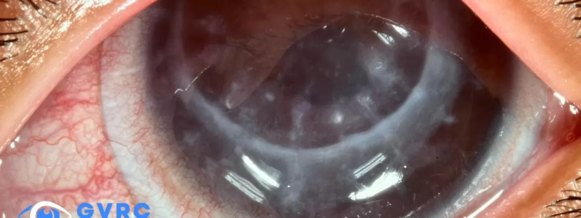
Scleral lenses may improve vision for selected people with Fuchs endothelial corneal dystrophy, but they do not treat the failing endothelial cells or remove corneal swelling. Because a scleral lens and its fluid reservoir can reduce oxygen reaching the cornea, candidacy requires particular caution when endothelial function is limited. Corneal thickness, edema, symptoms, lens fit, oxygen considerations, and the guidance of a corneal ophthalmologist all matter.
What Is Fuchs Endothelial Corneal Dystrophy?
Fuchs endothelial corneal dystrophy affects the endothelial cells lining the back of the cornea. These cells help pump excess fluid out of the cornea so it remains clear. Human endothelial cells have limited ability to replace themselves. As their number and function decline, fluid can accumulate and cause corneal edema.
For a broader overview of inherited dystrophies and acquired corneal changes, read Corneal Dystrophy and Degeneration.
Fuchs dystrophy often affects both eyes but may be asymmetric. Early disease may be found during an examination before a person notices symptoms. More advanced disease can cause blurred or fluctuating vision, glare, halos, reduced contrast, and light sensitivity. Vision may be worse after waking because less fluid evaporates from the corneal surface while the eyelids are closed. Persistent epithelial swelling can also cause painful blisters in advanced cases.
How Is Fuchs Dystrophy Evaluated?
Diagnosis is based on the history and eye examination. Slit-lamp findings may include corneal guttae, edema, haze, or epithelial changes. Pachymetry measures corneal thickness, while specular microscopy can provide information about endothelial cell density and morphology. Tomography, anterior-segment OCT, visual acuity, refraction, and glare symptoms may provide additional context.
A single cell count or thickness number does not determine treatment by itself. Measurements should be interpreted with symptoms, corneal clarity, the presence and pattern of edema, prior surgery, and change over time.
Can Scleral Lenses Help With Fuchs Dystrophy?
A scleral lens vaults the cornea and creates a new front optical surface. In a carefully selected eye, this may improve vision when irregular corneal optics, anterior corneal changes, or other refractive factors contribute to blur. A diagnostic lens trial can help determine whether the optical improvement is meaningful.
Not every reduction in vision from Fuchs dystrophy can be corrected with a lens. Light scatter from corneal edema or haze may continue even when the front optical surface is made smoother. If swelling or endothelial failure is the main reason for poor vision, a contact lens may offer limited benefit and does not address the underlying disease.
Why Oxygen and Corneal Edema Require Extra Caution
Every contact lens creates some resistance to oxygen reaching the cornea. A scleral lens adds a fluid reservoir beneath the lens, and lens thickness, material, clearance, wear time, tear exchange, and individual physiology can affect the response. Healthy corneas can show a small amount of swelling during scleral lens wear. An eye with compromised endothelial function may have less ability to remove additional fluid.
A 2026 systematic review and meta-analysis found small average and largely reversible corneal-thickness changes across the studied eyes during modern daytime scleral-lens wear, while still advising selective monitoring for higher-risk patients. Those averages do not establish safety for an individual eye with Fuchs dystrophy.
For this reason, Fuchs dystrophy is not a routine scleral-lens indication. The evaluation may include baseline corneal thickness, endothelial findings, edema, lens clearance, visual response, and repeat measurements after a controlled period of wear. A practitioner may consider a high-oxygen-permeable material, reduced lens or reservoir thickness where clinically appropriate, limited wear, or a different form of correction. These adjustments do not make every eye safe for scleral lens wear.
Increasing morning blur, persistent haze, halos, pain, redness, reduced wearing time, or vision that does not recover after lens removal should be reported promptly. Patients should follow the wear schedule and monitoring plan prescribed for their own eyes.
What Scleral Lenses Can and Cannot Do
- They may improve optical quality: A smooth lens surface can mask some irregular astigmatism and refractive distortion.
- They may provide a fluid reservoir: This can support the ocular surface in selected conditions, but it is not a treatment for endothelial pump failure.
- They do not restore endothelial cells: A lens cannot reverse Fuchs dystrophy or stop endothelial cell loss.
- They do not remove established edema: Corneal swelling may continue to blur vision and may worsen when oxygen delivery is insufficient.
- They do not replace corneal specialist care: Medical or surgical management may be needed when the disease becomes visually significant.
How Dr. Boshnick Evaluates a Fuchs Patient for Scleral Lenses
Dr. Edward Boshnick evaluates whether reduced vision appears optically correctable and whether a specialty lens can be worn with an acceptable physiological response. The process may include reviewing symptoms at different times of day, prior corneal or cataract surgery, current correction, ocular surface findings, corneal thickness, endothelial information, and the on-eye response to a diagnostic lens.
Fit assessment includes clearance, landing, centration, movement, comfort, over-refraction, and changes during wear. A corneal ophthalmologist remains the appropriate professional to diagnose and manage endothelial disease and to determine whether medical treatment or surgery is indicated. Care should be coordinated when edema is present, measurements are worsening, or a corneal procedure is being considered.
Other Treatment Options for Fuchs Dystrophy
Treatment depends on disease severity and the cause of the visual symptoms. Observation may be appropriate in mild disease. Hypertonic saline drops or ointment may temporarily reduce epithelial edema for some patients, but they do not restore endothelial cells or cure the condition. Prescription recommendations should come from the treating eye-care professional.
When corneal edema causes significant visual impairment or pain, a corneal surgeon may discuss endothelial keratoplasty. Descemet membrane endothelial keratoplasty, or DMEK, replaces the diseased endothelial layer and Descemet membrane. Descemet stripping automated endothelial keratoplasty, or DSAEK/DSEK, uses a somewhat thicker donor graft. Other approaches may be considered in selected cases. Each option has candidacy requirements, benefits, risks, and postoperative follow-up needs.
Can Scleral Lenses Be Used After Endothelial Keratoplasty?
Some patients need specialty optical correction after DMEK, DSAEK/DSEK, or another corneal procedure because of residual astigmatism, irregularity, scarring, or other factors. A post-surgical scleral lens evaluation requires clearance from the corneal surgeon and attention to graft attachment, endothelial function, corneal thickness, incisions, ocular surface health, oxygen delivery, and ongoing transplant monitoring.
A lens that improves vision does not remove the need to watch for graft rejection or failure. Sudden redness, pain, light sensitivity, or decreased vision after corneal transplantation requires urgent contact with the treating surgeon.
Questions to Ask During an Evaluation
- Is my blurred vision primarily caused by edema, irregular optics, cataract, retinal disease, or a combination?
- Do my corneal thickness and endothelial findings make a contact lens trial reasonable?
- How will the cornea be checked before, during, and after scleral lens wear?
- What wear-time limits or symptoms should prompt lens removal?
- Should a corneal ophthalmologist evaluate me before a lens trial?
- If surgery is recommended, when should specialty lens fitting be reconsidered?
Frequently Asked Questions
Can you wear contact lenses with Fuchs dystrophy?
Sometimes, but not automatically. A corneal or scleral lens may improve vision when refractive error or irregular optics are a meaningful part of the blur. It will not correct blur caused primarily by edema, and reduced endothelial function can increase concern about lens-related swelling. Candidacy depends on the examination, baseline measurements, a controlled lens trial, and follow-up.
Can scleral lenses cure Fuchs dystrophy?
No. Scleral lenses do not restore endothelial cells or cure Fuchs dystrophy. They may improve optical quality in selected eyes, but the underlying endothelial disease still requires monitoring and, in some cases, medical or surgical care.
Are scleral lenses safe for everyone with Fuchs dystrophy?
No. Reduced endothelial function and corneal edema can increase concern about oxygen-related swelling during lens wear. Safety depends on the severity of disease, baseline measurements, lens design, wear response, and ongoing follow-up.
Why is vision with Fuchs dystrophy often worse in the morning?
The closed eyelids reduce evaporation from the corneal surface during sleep. When endothelial pump function is limited, more fluid may remain in the cornea after waking, causing temporary additional swelling, blur, glare, or haze.
Can a scleral lens replace DMEK or another corneal procedure?
No. A scleral lens can provide removable optical correction but cannot replace failing endothelial cells. When edema or endothelial failure is the main problem, a corneal ophthalmologist should determine whether a procedure is appropriate.
What symptoms should a Fuchs patient report promptly?
Promptly report new or increasing pain, redness, light sensitivity, sudden or persistent loss of vision, corneal clouding, or vision that does not recover after lens removal. Transplant patients should follow their surgeon’s urgent-care instructions.
Request a Specialty Lens Evaluation
If Fuchs dystrophy or prior corneal surgery has left you with visual concerns that may be optically correctable, request an evaluation with Eye Freedom. A lens trial is considered only after the corneal health and likely cause of the symptoms are reviewed.
Medical Sources
- National Eye Institute: Corneal dystrophies
- American Academy of Ophthalmology EyeWiki: Fuchs endothelial dystrophy
- Systematic review and meta-analysis: Corneal responses to scleral lens wear
- Peer-reviewed study: Corneal optical changes associated with induced edema in Fuchs dystrophy
- Peer-reviewed study: Corneal abnormalities across Fuchs dystrophy severity
- Peer-reviewed study: Corneal edema before DMEK in Fuchs dystrophy
- American Academy of Ophthalmology patient guide: Fuchs dystrophy
This article is educational and does not provide a diagnosis or an individualized recommendation for contact lens wear, medication, or surgery.
