
Why Less Than 0.1% of Keratoconus Patients I Treat Need Surgery
Introduction
Keratoconus is a progressive eye condition characterized by the thinning and bulging of the cornea, the clear front surface of the eye, into a cone-like shape. This distortion of the cornea leads to blurred vision and light sensitivity, often impacting an individual’s quality of life. Traditionally, the treatment for keratoconus has been heavily reliant on corrective eyewear in the early stages and invasive surgical procedures in more advanced cases. These surgical interventions, while effective, carry potential risks and complications, and recovery can be a lengthy process.
However, in my practice, I’ve observed a remarkable trend: less than 0.1% of keratoconus patients I treat need surgery. This statement may seem surprising given the conventional treatment methods for this condition, but it’s a testament to the effectiveness of the innovative approaches we’ve adopted. In the forthcoming sections, we’ll delve into why such a small percentage of my keratoconus patients require surgical intervention and how our treatment methods are changing the narrative for individuals living with this condition.
Understanding Keratoconus
Keratoconus is an eye condition that primarily affects the cornea, the clear, dome-shaped front surface of the eye. In a healthy eye, the cornea is round and evenly curved. However, in an eye affected by keratoconus, the cornea progressively thins and bulges outward into a cone-like shape. This irregular shape distorts the light entering the eye, leading to blurred and distorted vision.
The exact cause of keratoconus is still unknown, but it’s believed to be a combination of genetic and environmental factors. Some research suggests that keratoconus may be linked to certain allergic conditions like asthma and eczema, and it may also be associated with chronic eye rubbing. It typically begins in the teenage years and progresses into the mid-30s.
Symptoms of keratoconus vary depending on the stage of the condition. In the early stages, individuals may experience slight blurring and distortion of vision, increased sensitivity to light, and frequent changes in eyeglass prescription. As the condition progresses, symptoms may include more pronounced vision distortion, increased astigmatism or nearsightedness, and inability to wear contact lenses due to discomfort.
The impact of keratoconus on a patient’s life can be significant. The progressive vision distortion can affect daily activities like reading, driving, and recognizing faces. It can also impact a person’s emotional well-being, leading to frustration and anxiety about the future. The traditional treatment approach, which often leads to surgery, can add to this anxiety.
However, the narrative doesn’t have to be this way. As we’ll explore in the next sections, less than 0.1% of keratoconus patients I treat need surgery, thanks to our innovative treatment methods.
Traditional Treatment Methods
The traditional treatment methods for keratoconus are largely dependent on the stage of the condition. In the early stages, glasses or soft contact lenses may be used to correct the mild astigmatism and nearsightedness caused by the initial corneal distortion. As the condition progresses and the cornea becomes more irregular, these methods become less effective.
Rigid gas-permeable (RGP) contact lenses are often the next line of treatment. These lenses maintain their shape, thereby providing a smooth refractive surface in front of the cornea. However, fitting these lenses can be challenging due to the irregular shape of the cornea, and some patients find them uncomfortable to wear.
When contact lenses are no longer a viable option, surgical interventions are considered. Corneal cross-linking is a procedure used to slow or halt the progression of keratoconus. It involves applying vitamin B2 drops to the cornea and then activating them with UV light to strengthen the corneal fibers.
In more severe cases, a corneal transplant may be necessary. This involves replacing the distorted cornea with healthy donor tissue. While corneal transplants can improve vision, they also carry potential risks and complications, such as rejection of the donor cornea, infection, and complications from long-term steroid use.
It’s important to note that while these traditional treatment methods can be effective, they also have limitations and potential downsides. This is why the fact that less than 0.1% of keratoconus patients I treat need surgery is so significant. In the following sections, we’ll explore the innovative treatment methods that make this possible.
My Approach to Treating Keratoconus
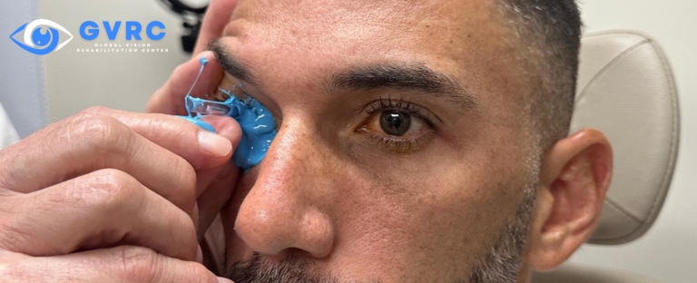
In my practice, I’ve adopted a unique approach to treating keratoconus that significantly reduces the need for surgical intervention. Central to this approach is the use of scleral lenses, a specialized type of contact lens that offers numerous benefits for keratoconus patients.
Scleral lenses are large-diameter gas permeable lenses that rest on the sclera, the white part of the eye, and vault over the entire corneal surface. This design allows for a smooth optical surface to correct vision distorted by keratoconus, while the space between the back surface of the lens and the cornea acts as a reservoir for fluid, providing continuous hydration that enhances comfort.
Unlike traditional contact lenses, scleral lenses are highly customizable and can be tailored to match the exact contours of the individual’s eye. This ensures a more comfortable fit, making them a viable option for patients who have previously struggled with lens discomfort.
My approach to treating keratoconus with scleral lenses differs significantly from traditional methods. Instead of merely correcting vision distortion or resorting to surgery when lenses fail, I focus on providing a non-surgical solution that offers improved vision comfort and quality of life. This approach has proven to be highly effective, with less than 0.1% of my keratoconus patients requiring surgery.
In the next section, we’ll delve deeper into why such a small percentage of my patients require surgical intervention and how scleral lenses are changing the lives of individuals living with keratoconus.
Why Less Than 0.1% of My Patients Need Surgery
The success of my approach to treating keratoconus is reflected in the fact that less than 0.1% of my patients require surgery. This is a significant departure from traditional treatment methods, where surgery is often the final resort when other options fail.
The key to this success lies in the use of scleral lenses. These lenses offer a non-surgical solution to the vision distortion caused by keratoconus. By creating a smooth optical surface over the irregular cornea, scleral lenses effectively correct vision while providing a comfortable fit. The customization of these lenses to each individual’s eye ensures optimal performance and patient satisfaction.
Moreover, the continuous hydration provided by the fluid reservoir between the lens and the cornea enhances comfort, making scleral lenses a viable long-term solution for many keratoconus patients. This eliminates the need for more invasive procedures, such as corneal transplants, which carry potential risks and complications.
To illustrate the effectiveness of this approach, consider the case of one of my patients, John. John was diagnosed with keratoconus in his early twenties and struggled with traditional contact lenses. His vision continued to deteriorate, and he was facing the prospect of a corneal transplant. However, after being fitted with scleral lenses, John’s vision improved significantly, and he was able to resume his normal activities without the need for surgery.
John’s story is just one of many. Time and again, I’ve seen how scleral lenses can transform the lives of keratoconus patients, providing them with improved vision and a better quality of life. This innovative approach to treatment is why less than 0.1% of my keratoconus patients require surgery, a testament to the power of non-surgical solutions in managing this condition.
Conclusion
In conclusion, keratoconus is a progressive eye condition that has traditionally been treated with corrective eyewear, rigid gas-permeable lenses, and ultimately, surgical interventions. However, these methods, while effective to some extent, have their limitations and potential downsides.
In contrast, my approach to treating keratoconus centers around the use of scleral lenses, a non-surgical solution that offers improved vision and comfort for patients. These lenses are highly customizable, providing a smooth optical surface over the irregular cornea and a comfortable fit. The success of this approach is evident in the fact that less than 0.1% of my keratoconus patients require surgery.
This article has explored the nature of keratoconus, traditional treatment methods, and my unique approach to managing this condition. The key takeaway is that surgery is not the only option for keratoconus patients. Non-surgical solutions, such as scleral lenses, can offer effective treatment and a better quality of life.
If you or a loved one are living with keratoconus, I encourage you to explore these non-surgical treatment options. With the right approach, it’s possible to manage this condition effectively and enjoy a life with clearer vision.