7800 SW 87 AVE SUITE B-270 MIAMI, FLORIDA 33173

What Are Wavefront-Guided Scleral Lenses?

Wavefront-guided scleral lenses are custom scleral lenses with optics designed from measurements of an individual’s remaining optical distortions while a stable lens is on the eye. They may be considered when glasses or a conventional scleral lens improve visual acuity but glare, halos, ghosting, starbursts, reduced contrast, or poor night vision remain.

The lens still must vault the cornea, land appropriately on the sclera, remain stable, and support ocular health. Wavefront-guided optics are not automatically better for every patient, and they cannot guarantee elimination of a symptom.

Higher-Order Aberrations and Visual Quality

Standard prescriptions correct lower-order refractive errors such as nearsightedness, farsightedness, and regular astigmatism. Higher-order aberrations are more complex distortions in the eye’s optical system. Examples include coma, trefoil, and spherical aberration.

These aberrations can affect contrast and image quality even when a patient can read small letters on a high-contrast eye chart. Symptoms can include monocular ghost images, halos around lights, streaking, glare, starbursts, or blur that becomes more noticeable in dim illumination.

Symptoms alone do not prove that higher-order aberrations are the cause. Tear-film instability, lens deposits, residual prescription, lens flexure, decentration, rotation, cataract, retinal disease, or other problems may produce similar complaints and must be evaluated.

How Wavefront-Guided Optics Differ From Standard Scleral Lenses

A conventional custom scleral lens creates a smooth anterior optical surface and a fluid reservoir over an irregular cornea. This alone can reduce many corneal aberrations and provide useful vision for numerous patients.

Wavefront-guided design adds a customized front-surface optical pattern intended to address selected residual aberrations measured through the eye and lens together. Because that correction must remain aligned with the eye, an appropriately centered and rotationally stable scleral lens fit is the foundation of the process.

If the lens moves, rotates, decenters, flexes, or changes position after settling, the customized optics may not remain aligned as intended. Fit, ocular surface condition, tear-film behavior, and ordinary prescription refinement therefore come before wavefront customization.

Who May Be Evaluated?

Dr. Edward Boshnick may evaluate wavefront-guided scleral optics for selected patients with measurable residual aberrations and a sufficiently stable scleral lens platform. Examples may include people with:

  • keratoconus or pellucid marginal degeneration;
  • corneal ectasia after refractive surgery;
  • post-LASIK or post-RK irregularity;
  • corneal scarring or a prior corneal transplant;
  • irregular astigmatism; or
  • persistent glare, halos, ghosting, or reduced contrast despite an otherwise appropriate correction.

Candidacy depends on examination and measurement. Many patients do not need wavefront-guided optics and may obtain an appropriate result with a standard custom scleral lens.

How Evaluation and Fitting Work

  1. Identify the cause of the symptoms: Dr. Boshnick reviews the diagnosis, prior procedures, current correction, ocular surface, tear film, lens condition, and visual goals.
  2. Establish an appropriate scleral lens fit: Clearance, limbal relationship, scleral landing, centration, rotation, settling, and tissue response are evaluated.
  3. Refine ordinary optics: Sphere, cylinder, axis, over-refraction, lens flexure, and other correctable factors are addressed first.
  4. Measure residual aberrations: Wavefront aberrometry is performed through the stable lens when the readings are repeatable and clinically meaningful.
  5. Design and evaluate the customized optics: The manufactured lens is assessed for alignment, vision, comfort, and ocular health.
  6. Follow up during real-world wear: The fit and optics may require refinement after settling and adaptation.

Bring current and prior lenses, available lens parameters, corneal imaging, surgical records, and notes about when symptoms are most noticeable.

WHAT THE MEASUREMENTS SHOW

Wavefront Measurements Used in Scleral Lens Evaluation

Wavefront map used in advanced scleral lens planning

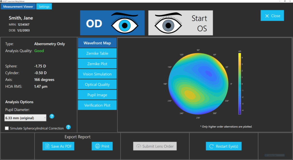
Wavefront Map

A wavefront map represents how light passing through the eye differs from an ideal optical system. Dr. Boshnick interprets the pattern together with refraction, lens position, pupil size, corneal findings, and the patient’s symptoms.

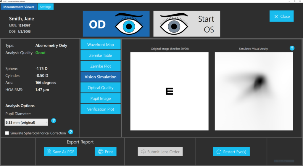
Simulated Vision

A simulated image can help explain how measured optical distortions may affect visual quality. It is an educational representation, not a guarantee of what a patient will see with a particular lens.

Wavefront map used in advanced scleral lens planning

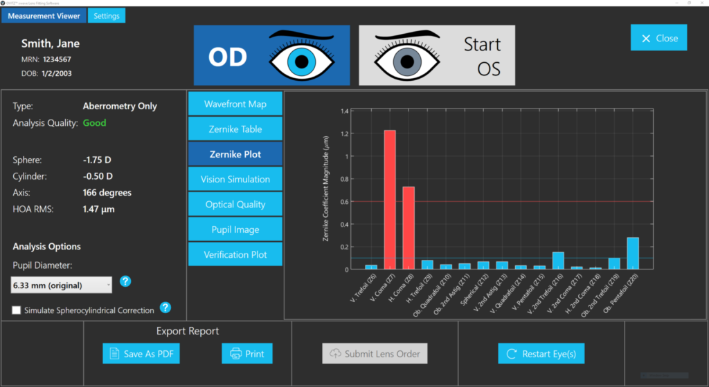
Zernike Terms

Zernike terms are a mathematical way to describe optical aberrations such as coma, trefoil, and spherical aberration. The measurement must be repeatable and interpreted in the context of the full examination.

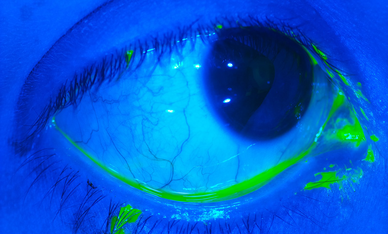
Ocular and Lens Imaging

High-resolution imaging helps document lens position, surface condition, fluorescein patterns, and ocular findings. Imaging complements, but does not replace, direct clinical assessment.

Wavefront aberrometer technology for specialty lens evaluation

Dr. Boshnick's Clinical Approach

Dr. Edward Boshnick evaluates complex corneal and post-surgical cases by separating fitting, ocular surface, and optical causes of reduced visual quality. Wavefront data are used only when they answer a specific clinical question and can be incorporated into a stable lens design.

That sequence matters: a sophisticated optical design cannot compensate for an unhealthy ocular surface, unstable lens position, incorrect filling or care, or a condition outside the cornea and anterior optical system.

What Wavefront-Guided Lenses Can and Cannot Do

Research has shown that wavefront-guided scleral lenses can reduce measured residual aberrations and improve selected measures of visual performance in some eyes with keratoconus or irregular corneal astigmatism. Results vary, and high-contrast acuity may not fully describe a patient’s experience.

These lenses do not reverse corneal disease, stop keratoconus progression, or guarantee normal night vision. New pain, pronounced redness, discharge, sudden light sensitivity, or an abrupt reduction in vision requires prompt professional attention.

Read about wavefront aberrometry, the WaveDyn Vision Analyzer, or the broader scleral lens evaluation process.

Medical sources

This page is educational and does not replace an eye examination, diagnosis, or individualized treatment plan.

Frequently Asked Questions

It is a custom scleral lens with front-surface optics designed from measurements of selected residual aberrations through a stable lens on the eye. The physical lens fit and standard prescription must be established first.
No. Many patients obtain appropriate vision with conventional custom scleral lenses. Wavefront-guided optics are considered when repeatable residual aberrations are limiting visual quality and the lens can remain sufficiently stable.
They may reduce these symptoms for selected patients when higher-order aberrations are an important cause. Similar symptoms can come from tear-film, lens, cataract, retinal, or other problems, so examination is necessary.
The customized optical pattern must stay aligned with the measured aberrations. Lens decentration, rotation, movement, flexure, or settling can change that alignment and reduce the intended effect.
No. They are used for optical rehabilitation. Corneal cross-linking is a separate procedure commonly considered to reduce progression risk in appropriate patients.
Bring current and prior lenses, available lens parameters, corneal imaging, surgical records, a medication and eye-drop list, and notes about lighting or activities that make the symptoms better or worse.

Schedule Your Consultation with
Dr. Boshnick