

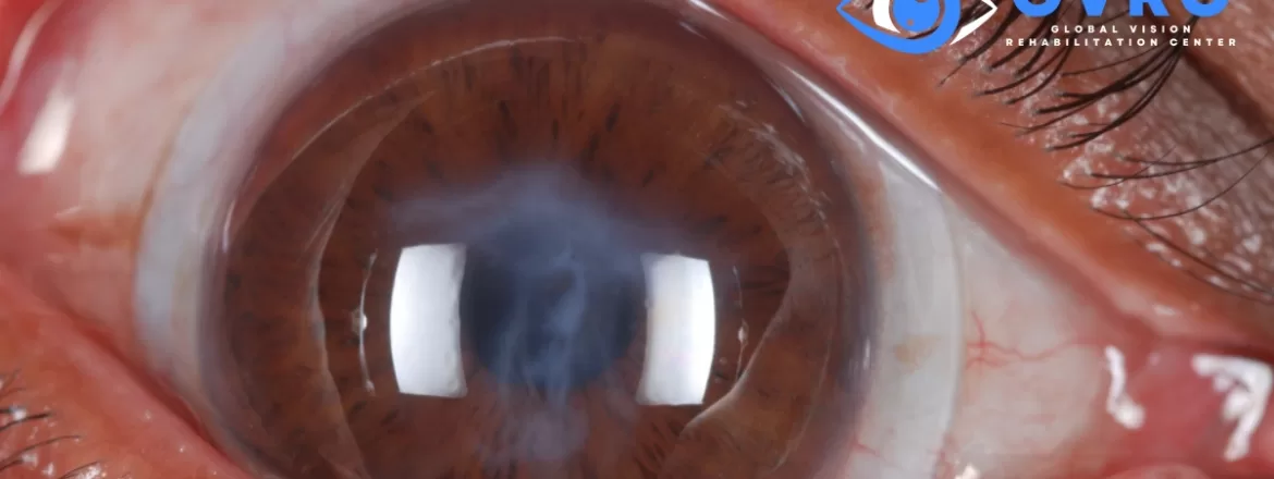
Non-surgical keratoconus treatment usually has two separate goals: improve vision and monitor or limit progression. Glasses and specialty contact lenses can help a person see more clearly, but they do not strengthen the cornea or stop keratoconus from changing. Corneal cross-linking may be recommended by a corneal ophthalmologist when progression is documented. The right plan depends on corneal shape, stability, scarring, ocular surface health, age, and the vision achieved with each form of correction.
Key Takeaways
- Mild keratoconus may be corrected with glasses or soft contact lenses.
- Rigid, hybrid, or scleral lenses may provide better optical correction when the cornea becomes irregular.
- Scleral lenses improve vision; they do not stop keratoconus progression.
- Corneal cross-linking and specialty lenses serve different purposes and may both be part of care.
- Corneal transplant surgery is generally considered only when less invasive options do not provide adequate function or when significant scarring or other complications are present.
Can Keratoconus Be Managed Without Corneal Transplant Surgery?
Many people with keratoconus can achieve useful vision without a corneal transplant. Depending on the stage of the condition, management may include glasses, soft lenses, rigid corneal lenses, hybrid lenses, or custom scleral lenses for keratoconus. Regular corneal imaging is also important because a lens can improve vision even while the underlying cornea is changing.
Avoiding transplant surgery is not a promise or the only measure of successful care. Some patients need a corneal procedure to address documented progression, severe scarring, thinning, hydrops, or vision that remains inadequate with contact lenses. An examination is needed to separate optical rehabilitation from treatment of the corneal disease itself.
What Is Keratoconus?
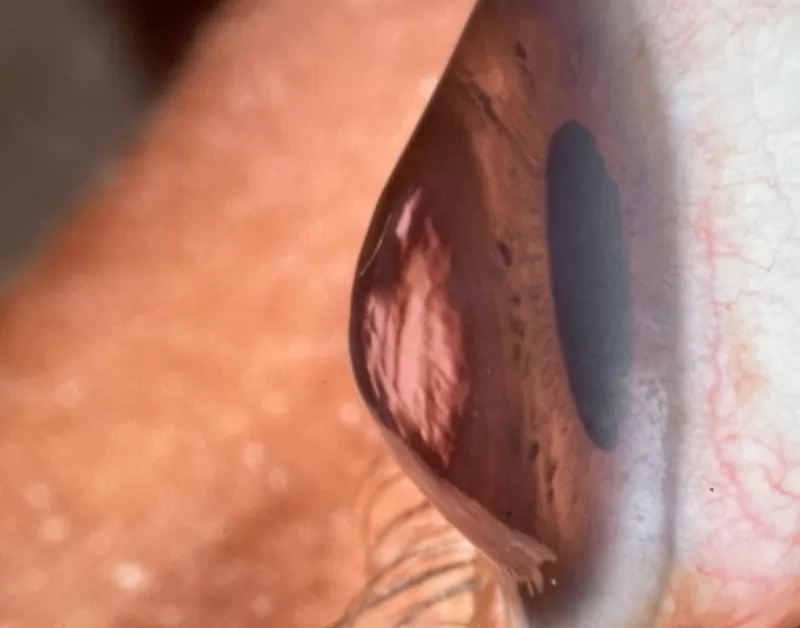
Keratoconus is a corneal ectasia in which the cornea becomes thinner and develops a steeper, more irregular shape. Because the cornea provides much of the eye’s focusing power, this change can produce irregular astigmatism, increasing nearsightedness, ghosting, glare, halos, multiple images, and reduced contrast.
Early symptoms may resemble an ordinary prescription change. As irregularity increases, glasses may correct only part of the blur because a standard lens cannot fully neutralize an uneven corneal surface. Corneal topography or tomography, refraction, slit-lamp examination, and corneal-thickness measurements help establish the diagnosis and monitor change.
Non-Surgical Ways to Improve Vision With Keratoconus
Glasses and Soft Contact Lenses
Glasses or conventional soft lenses may provide acceptable vision in early or relatively regular keratoconus. They are familiar and simple to use, but their ability to correct vision can decrease as irregular astigmatism becomes more pronounced.
Rigid Corneal and Hybrid Lenses
A rigid gas-permeable corneal lens creates a smoother front optical surface than the irregular cornea beneath it. Hybrid designs combine a rigid center with a soft skirt. These options can work well for selected eyes, although lens stability, centration, comfort, and corneal shape all affect the result.
Scleral Lenses
A scleral lens is a large-diameter gas-permeable lens designed to vault the cornea and land on tissue over the sclera. The space between the lens and cornea is filled with preservative-free saline before insertion. The lens’s front optical surface can mask some corneal irregularity and may provide clearer, more stable vision for selected patients.
Because the lens does not rest on the cone in the same way as a smaller corneal lens, it may also offer a useful fitting option when a corneal lens is unstable or uncomfortable. Results vary, and successful wear depends on a carefully evaluated fit, adequate oxygen delivery, handling ability, lens care, ocular surface health, and follow-up.
What Scleral Lenses Can and Cannot Do
Scleral lenses are used for visual rehabilitation. They may reduce blur, ghosting, and fluctuations related to irregular corneal optics. Some patients also value the fluid reservoir when ocular surface disease is present, although a keratoconus fit must still be assessed individually.
Scleral lenses do not reverse corneal thinning, permanently reshape the cornea, or stop keratoconus progression. Clear vision through a lens does not prove that the condition is stable. Follow-up corneal imaging remains important, particularly in younger patients and anyone with changing vision or prescription.
Cross-Linking and Scleral Lenses Have Different Jobs
Corneal collagen cross-linking uses riboflavin and ultraviolet light to increase biomechanical stability in the cornea. A corneal ophthalmologist may consider it when keratoconus is progressive and the eye meets the clinical requirements for a specific protocol. Cross-linking is intended to slow or stabilize progression; it does not guarantee that the cornea will become regular or that glasses will provide adequate vision afterward.
Scleral lenses are intended to improve optical function. A person may therefore be evaluated for cross-linking to address progression and later use glasses or specialty contact lenses for vision. Eye Freedom does not present a specialty lens as a substitute for indicated corneal treatment. When progression or a procedure is a concern, Dr. Edward Boshnick coordinates or recommends evaluation with an appropriate corneal specialist.
How Dr. Boshnick Evaluates a Keratoconus Lens Fit
Dr. Boshnick’s specialty-lens evaluation begins with the patient’s visual history, current correction, symptoms, previous lens experience, and treatment history. Corneal shape and ocular surface findings help guide which lens designs should be evaluated. A diagnostic lens is assessed on the eye rather than selected from a diagnosis alone.
During fitting and follow-up, clinically relevant factors may include central and limbal clearance, landing alignment, centration, movement, visual acuity, over-refraction, comfort, removal findings, and changes that develop during wear. Some optically complex eyes may also be evaluated for wavefront-guided scleral lens technology, but not every patient needs or benefits from a wavefront-guided design.

Who May Be a Candidate for Scleral Lenses?
An evaluation may be reasonable when keratoconus causes inadequate or unstable vision with glasses or soft lenses, when a corneal rigid lens does not center or remain comfortable, or when the eye has additional optical or ocular surface complexity. Candidacy cannot be determined from a scan, prescription, or diagnosis alone.
Active inflammation, infection, severe handling difficulty, poor follow-up, certain endothelial or oxygen-related concerns, and other ocular findings may affect whether scleral lens wear is appropriate. Patients should also understand the daily filling, insertion, removal, cleaning, disinfection, and replacement requirements before proceeding.
When May Keratoconus Surgery Still Be Considered?
A corneal ophthalmologist may discuss surgical or procedural options when there is documented progression, substantial scarring, advanced thinning, recurrent complications, contact lens intolerance, or inadequate functional vision despite appropriate correction. Options differ by patient and may include cross-linking, intracorneal ring segments in selected cases, deep anterior lamellar keratoplasty, or penetrating keratoplasty.
Even after a procedure, some patients still need glasses or specialty contact lenses for their best vision. The decision should be based on corneal health, progression, functional needs, expected benefits, and risks rather than on a fixed claim that one option is always best.
Questions to Ask at a Keratoconus Evaluation
- Does my imaging show that the cornea is stable or changing?
- Is the immediate goal better vision, control of progression, or both?
- Which types of contact lenses are reasonable for my corneal shape and ocular surface?
- What should I expect during a scleral lens trial and follow-up?
- Do I need a corneal ophthalmology consultation for cross-linking or another procedure?
- Which symptoms should prompt an earlier examination?
Request a Keratoconus and Scleral Lens Evaluation
Eye Freedom evaluates patients who travel from across the United States and internationally for complex corneal and specialty contact lens care. An examination with Dr. Boshnick can determine whether a scleral lens trial is appropriate and whether additional corneal evaluation should be part of the plan.
Request an evaluation with Eye Freedom.
Frequently Asked Questions
What is the best non-surgical treatment for keratoconus?
There is no single best option for every patient. Glasses or soft lenses may be sufficient in mild disease, while rigid, hybrid, or scleral lenses may provide better vision with greater corneal irregularity. If progression is documented, a corneal specialist may recommend cross-linking. Optical correction and progression control are different parts of care.
Can scleral lenses stop keratoconus from getting worse?
No. Scleral lenses can improve vision by creating a smoother optical surface, but they do not stop corneal thinning or progression. Continued corneal monitoring remains important.
Can scleral lenses help me avoid a corneal transplant?
For some patients, specialty lenses provide adequate functional vision and a transplant is not needed at that time. They cannot eliminate the possibility of surgery in every case, particularly when severe scarring, thinning, intolerance, or other complications are present.
Is cross-linking the same as wearing a scleral lens?
No. Cross-linking is a corneal procedure intended to slow or stabilize progression in eligible eyes. A scleral lens is removable optical correction intended to improve vision. A patient may need one, both, or neither depending on the examination.
How often should keratoconus be monitored?
The interval depends on age, prior measurements, symptoms, and risk of progression. A clinician may recommend closer follow-up when the condition is newly diagnosed, changing, or present in a younger patient. Keep the schedule recommended by the professionals managing the cornea and contact lenses.
Medical Sources
- American Academy of Ophthalmology EyeWiki: Keratoconus
- U.S. Food and Drug Administration: Corneal cross-linking summary review
- Systematic review: Long-term effectiveness of scleral lenses in keratoconus
- Prospective study: Vision and quality of life with scleral lenses in keratoconus
- Review: Scleral lenses, visual acuity, and ocular aberrations in corneal ectasia
This article is educational and does not provide a diagnosis or an individualized treatment recommendation. Seek prompt eye care for sudden loss of vision, new pain, marked light sensitivity, redness, or sudden corneal clouding.
