

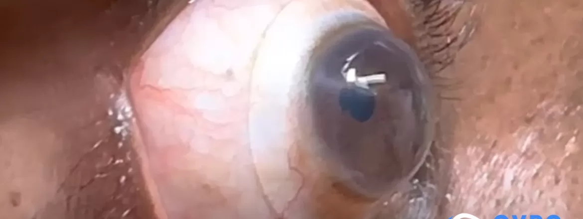
Common signs of keratoconus include distorted or blurred vision, ghost images, glare or halos, frequent prescription changes, and vision that glasses or standard soft contact lenses no longer correct well. These symptoms do not prove that someone has keratoconus, but they are reasons to schedule a comprehensive eye examination with corneal mapping.
Keratoconus changes the shape of the cornea, the clear front surface of the eye. As the cornea becomes thinner and more irregular, light may no longer focus as a single clear image. Symptoms can begin subtly, affect the two eyes differently, and change over time.
Five Signs That May Suggest Keratoconus
1. Blurred or Distorted Vision That Keeps Changing
Early keratoconus may resemble ordinary nearsightedness or astigmatism. The difference is that the corneal shape can become increasingly irregular, so a new eyeglass prescription may help temporarily but fail to provide consistently sharp vision.
Words may look smeared, straight lines may appear slightly bent, or fine details may remain unclear even with correction. One eye may be affected more than the other.
2. Ghost Images or Double Vision in One Eye
Some people see a faint second image, shadow, or multiple outlines around letters and lights. If the symptom remains when the other eye is covered, it is called monocular diplopia. Irregular corneal optics can cause this type of ghosting, although other conditions can produce similar symptoms.
A routine vision screening alone may not identify why the image is distorted. Corneal measurements help determine whether the cause is keratoconus, another form of irregular astigmatism, tear-film instability, or a different eye condition.
3. Increasing Glare, Halos, or Difficulty Seeing at Night
An irregular cornea can scatter light rather than focusing it cleanly. Headlights, streetlights, and bright signs may develop halos, starbursts, streaks, or multiple images. Night driving may become more difficult even when daytime vision seems acceptable.
Glare and halos are common symptoms with many possible causes. Their presence is a reason for evaluation, not a diagnosis by itself.
4. Frequent Prescription Changes or Increasing Astigmatism
A prescription that changes repeatedly, especially with increasing or irregular astigmatism, can be a clue that the corneal shape should be measured. Younger patients and people with a family history of keratoconus may need closer assessment when these changes occur.
Bring prior eyeglass and contact lens prescriptions to the examination when possible. Comparing them can help the eye-care team understand the pattern and rate of change.
5. Glasses or Standard Soft Contacts No Longer Provide Stable Vision
Glasses and conventional soft lenses work best when the eye’s focusing surface is relatively regular. As keratoconus becomes more irregular, those options may leave residual blur, ghosting, or fluctuating clarity. A soft lens may also move unpredictably or fail to mask the corneal distortion.
This does not mean every person with these difficulties needs a scleral lens. Depending on the eye, options may include updated glasses, specialty soft lenses, corneal rigid lenses, hybrid lenses, or custom scleral lenses.
When a Vision Change Needs Prompt Attention
Seek prompt eye care for a sudden drop in vision, new eye pain, pronounced redness, marked light sensitivity, or sudden corneal clouding. In a person with keratoconus, rapid clouding can be associated with acute corneal hydrops and should not wait for a routine lens appointment.
How Keratoconus Is Confirmed
Symptoms alone cannot confirm keratoconus. An examination may include refraction, slit-lamp evaluation, and measurements of corneal shape and thickness. Corneal topography maps surface curvature, while tomography can evaluate the front and back of the cornea and its thickness distribution.
Comparison over time matters because a single examination shows the present shape, while serial measurements help identify progression. The Eye Freedom keratoconus guide explains diagnosis, monitoring, and the two separate goals of care.
Where Scleral Lenses May Fit
A custom scleral lens rests on the sclera, the white part of the eye, and vaults over the irregular cornea. Preservative-free saline fills the space between the lens and cornea. The lens and fluid reservoir create a regular front optical surface that may reduce blur and distortion caused by irregular astigmatism.
Scleral lenses can improve vision for selected patients, but they do not cure keratoconus, strengthen the cornea, or stop progression. Corneal cross-linking has a different purpose and may be considered by a corneal specialist when progression is present or likely.
Dr. Edward Boshnick evaluates corneal shape, ocular surface health, prior correction, diagnostic lens behavior, clearance, alignment, comfort, and visual quality before recommending a lens design. Read more about scleral lenses for keratoconus and the non-surgical keratoconus treatment options.
What to Bring to a Keratoconus Evaluation
- recent and older eyeglass or contact lens prescriptions;
- prior corneal topography, tomography, or cross-linking records;
- current lenses, cases, plungers, and care products;
- a list of symptoms and when they are most noticeable; and
- information about family history, allergies, and eye rubbing.
A diagnostic lens trial may help determine how much visual improvement is possible before a custom design is finalized.
Frequently Asked Questions
What is usually the first sign of keratoconus?
There is no single first sign for everyone. Blurred or distorted vision, increasing astigmatism, ghosting, glare, and frequent prescription changes are common reasons for further corneal evaluation.
Can an ordinary eye exam detect keratoconus?
An examination may reveal clues, but corneal topography or tomography is often important for detecting and documenting the irregular shape, particularly when changes are subtle.
Does having keratoconus mean I need scleral lenses?
No. The appropriate correction depends on the corneal shape, stage, symptoms, comfort, and visual needs. Some people do well with glasses or other contact lens designs; others may benefit from scleral lenses.
Can scleral lenses stop keratoconus from progressing?
No. Scleral lenses are used for vision rehabilitation and, in some cases, ocular surface protection. They do not stop progression. Cross-linking and other progression decisions require a separate corneal evaluation.
When should I request an evaluation?
Consider an evaluation when vision changes repeatedly, glasses no longer provide stable clarity, glare or ghosting interferes with daily activities, or keratoconus has already been diagnosed and current correction is not working well. Use the Eye Freedom contact form to request an appointment.
Medical Sources
This article is educational and does not diagnose keratoconus or recommend an individualized treatment.
